 CTICases
Type A Aortic Dissection with Acute Bilateral Lower Extremity Paralysis and Retroperitoneal Hematoma
 ▶ Watch on YouTube — age-restricted, cannot embed here
[...]. Type A Aortic Dissection with Acute Bilateral Lower Extremity Paralysis and Retroperitoneal Hematoma. CTICases. Published October 4, 2015. https://cticases.com/c/pywnd
Imaging findings
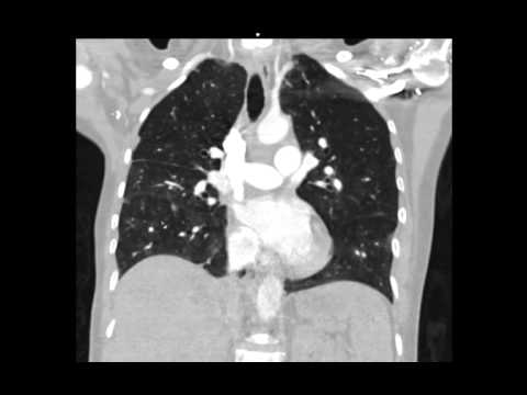
A middle-aged man presented with acute onset bilateral lower extremity paralysis. CT revealed a type A aortic dissection, potentially involving the aortic root (evidenced by blood along the pulmonary artery). The dissection extended distally, with a flap identified at the celiac axis and into the SMA. Critically, arterial and delayed venous phase imaging showed complete loss of opacification in the infrarenal aorta and iliacs, leading to lower extremity devascularization. A retroperitoneal hematoma suggested an aortic or iliac rupture.
Key takeaways
Type A aortic dissection can present with acute bilateral lower extremity paralysis due to abrupt occlusion of the infrarenal aorta, a severe manifestation resembling Leriche syndrome. This case demonstrates the extensive reach of aortic dissection and its potential for catastrophic complications, including lower limb compartment syndrome, rhabdomyolysis, and multiorgan failure, emphasizing the need for prompt recognition and intervention.
AI-assisted summary — may contain errors. Verify against the source video. Learn more
More from this webinar
- Pulmonary Tumor Thrombotic Microangiopathy in Prostate CancerNeoplastic
- Pulmonary Tumor Thrombotic Microangiopathy in Metastatic Breast CancerNeoplastic
- Spontaneous Auto-Embolization of a Pulmonary Arteriovenous Malformation (AVM)Vascular
- Iatrogenic Diaphragmatic Herniation of Mesenteric FatIatrogenic
- Metastatic Renal Cell Carcinoma Presenting as Large Pleural Effusion and Pleural MetastasesNeoplastic
- Coronary Artery Aneurysms and Occlusion in Eosinophilic Granulomatosis with Polyangiitis (EGPA) VariantAutoimmune
- Microvascular Plug (MVP) Embolization of Pulmonary Arteriovenous Malformations (AVMs)Iatrogenic
- Sclerotic Bone Metastases from Breast Cancer Mimicking Pulmonary NodulesNeoplastic
See all cases from October 4, 2015 →
Related Vascular cases
- Mycotic Aneurysm (from Enterococcus Faecalis Endocarditis)
- Left Ventricular Pseudoaneurysm
- Chronic Type B Aortic Dissection with False Lumen Aneurysm
- Denver Shunt-Associated Fibrin Sheath and Right Atrial Thrombosis
- Extensive Pulmonary Embolism with Cavitating Infarction
- Transpleural Systemic-to-Pulmonary Venous Collateral in Central Venous Obstruction