 CTICases
Pulmonary Tumor Thrombotic Microangiopathy in Metastatic Breast Cancer
 ▶ Watch on YouTube — age-restricted, cannot embed here
[...]. Pulmonary Tumor Thrombotic Microangiopathy in Metastatic Breast Cancer. CTICases. Published October 4, 2015. https://cticases.com/c/p2p8p
Imaging findings
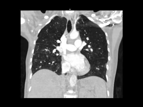
A second patient with metastatic breast cancer and pulmonary hypertension showed similarly enlarged right heart chambers with septal straightening on CT. CTPA (rather than a V/Q scan) demonstrated a mosaic attenuation pattern with patchy areas of relative hypo- and hyperperfusion of the pulmonary vasculature, along with small nodules possibly representing intravascular tumor.
Key takeaways
Mosaic attenuation on CTPA can be the CT correlate of the perfusion defects seen with pulmonary tumor thrombotic microangiopathy on V/Q scanning, reflecting the same underlying pathology of tumor emboli plus fibrocellular intimal proliferation in small pulmonary vessels. This patient also died soon after presentation, reinforcing PTTM's grave prognosis.
AI-assisted summary — may contain errors. Verify against the source video. Learn more
More from this webinar
- Pulmonary Tumor Thrombotic Microangiopathy in Prostate CancerNeoplastic
- Spontaneous Auto-Embolization of a Pulmonary Arteriovenous Malformation (AVM)Vascular
- Iatrogenic Diaphragmatic Herniation of Mesenteric FatIatrogenic
- Metastatic Renal Cell Carcinoma Presenting as Large Pleural Effusion and Pleural MetastasesNeoplastic
- Coronary Artery Aneurysms and Occlusion in Eosinophilic Granulomatosis with Polyangiitis (EGPA) VariantAutoimmune
- Microvascular Plug (MVP) Embolization of Pulmonary Arteriovenous Malformations (AVMs)Iatrogenic
- Type A Aortic Dissection with Acute Bilateral Lower Extremity Paralysis and Retroperitoneal HematomaVascular
- Sclerotic Bone Metastases from Breast Cancer Mimicking Pulmonary NodulesNeoplastic
See all cases from October 4, 2015 →