 CTICases
Williams Syndrome with Supravalvular Aortic Stenosis and Pulmonary Artery Narrowing
 ▶ Watch on YouTube — age-restricted, cannot embed here
[...]. Williams Syndrome with Supravalvular Aortic Stenosis and Pulmonary Artery Narrowing. CTICases. Published April 3, 2014. https://cticases.com/c/4up4k
Imaging findings
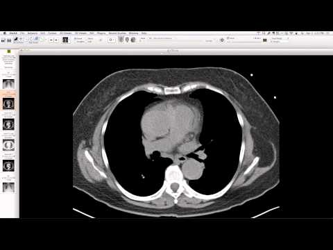
Diffuse narrowing of the entire supravalvular aorta from the sinotubular junction, measuring approximately 6-7 mm throughout, without normal tapering. Associated relatively mild narrowing of the right pulmonary artery, while the left pulmonary artery was normal.
Key takeaways
This is a genetically confirmed case of Williams syndrome, which is associated with diffuse supravalvular aortic stenosis and can involve pulmonary artery narrowing. While often suspected, true positive cases are relatively uncommon to encounter.
AI-assisted summary — may contain errors. Verify against the source video. Learn more
More from this webinar
- Alveolar Capillary Dysplasia with Unroofed Coronary Sinus (Right-to-Left Shunt)Congenital
- Iatrogenic Esophageal Rupture from Blakemore Tube PlacementIatrogenic
- Primum Atrial Septal Defect (ASD)Congenital
- Pulmonary Alveolar MicrolithiasisMetabolic
- Esophageal Varices Anastomosing with an Inferior Pulmonary VeinVascular
- Unroofed Coronary Sinus (Isolated Finding)Congenital
- Complex Congenital Anomalies: Right Aortic Arch with Aberrant Innominate Artery, Total Anomalous Pulmonary Venous Return of Left Lung, and Cor Triatriatum SinisterCongenital
- Unilateral Perched Facet at C7-T1 with ParaplegiaTrauma
See all cases from April 3, 2014 →
Related Congenital cases
- Repaired Tetralogy of Fallot with Left Isomerism, Azygos Continuation of IVC, and Aortic Stenosis
- Foramen of Morgagni Hernia (Incidental)
- Retroesophageal Right Vertebral Artery from Diverticulum of Kommerell (Aortic Arch Variant)
- Circumflex Aorta
- High Origin of the Right Coronary Artery
- Atrial Septal Defect