 CTICases
Unilateral Perched Facet at C7-T1 with Paraplegia
 ▶ Watch on YouTube — age-restricted, cannot embed here
[...]. Unilateral Perched Facet at C7-T1 with Paraplegia. CTICases. Published April 3, 2014. https://cticases.com/c/u5fc8
Imaging findings
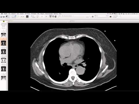
On chest radiograph, initial subtle widening of the superior mediastinum, without tracheal displacement. Separation of posterior elements at T1 and C7, with non-aligned spinous processes and facet separation. Follow-up radiographs show a larger hematoma and a "perched cassette" appearance. CT confirms a unilateral perched facet at C7-T1 and fracture of T1 posterior elements. A paravertebral hematoma is present, with no vascular injury.
Key takeaways
While perched facets are typically seen in the mid-cervical spine, this case demonstrates a unilateral perched facet at the cervicothoracic junction (C7-T1), a less common location that can still result in severe neurological deficits such as paraplegia. It serves as a good reminder to meticulously evaluate the upper thoracic spine on chest radiographs for subtle signs of trauma.
AI-assisted summary — may contain errors. Verify against the source video. Learn more
More from this webinar
- Williams Syndrome with Supravalvular Aortic Stenosis and Pulmonary Artery NarrowingCongenital
- Alveolar Capillary Dysplasia with Unroofed Coronary Sinus (Right-to-Left Shunt)Congenital
- Iatrogenic Esophageal Rupture from Blakemore Tube PlacementIatrogenic
- Primum Atrial Septal Defect (ASD)Congenital
- Pulmonary Alveolar MicrolithiasisMetabolic
- Esophageal Varices Anastomosing with an Inferior Pulmonary VeinVascular
- Unroofed Coronary Sinus (Isolated Finding)Congenital
- Complex Congenital Anomalies: Right Aortic Arch with Aberrant Innominate Artery, Total Anomalous Pulmonary Venous Return of Left Lung, and Cor Triatriatum SinisterCongenital
See all cases from April 3, 2014 →