 CTICases
Primum Atrial Septal Defect (ASD)
 ▶ Watch on YouTube — age-restricted, cannot embed here
[...]. Primum Atrial Septal Defect (ASD). CTICases. Published April 3, 2014. https://cticases.com/c/ph6qn
Imaging findings
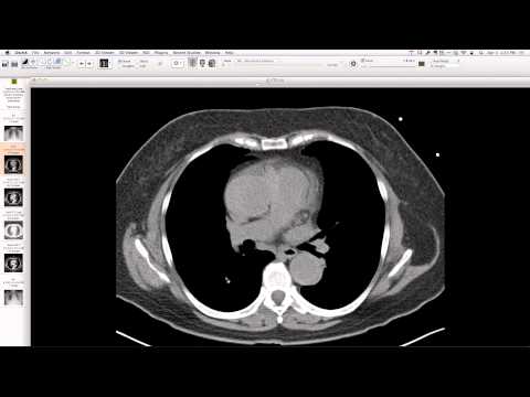
Large pulmonary arteries, a large primum atrial septal defect, and a single valve plane with some irregularity of the anterior leaflet of the mitral valve. Extensive left-to-right shunting across the ASD was observed, with a Qp:Qs ratio of approximately 2.8:1 to 3:1.
Key takeaways
Primum ASDs are a form of congenital heart disease, often associated with a cleft in the anterior leaflet of the mitral valve. Large shunts can lead to significant pulmonary overcirculation. It is notable for a patient to present with an undiagnosed large primum ASD in middle age.
AI-assisted summary — may contain errors. Verify against the source video. Learn more
More from this webinar
- Williams Syndrome with Supravalvular Aortic Stenosis and Pulmonary Artery NarrowingCongenital
- Alveolar Capillary Dysplasia with Unroofed Coronary Sinus (Right-to-Left Shunt)Congenital
- Iatrogenic Esophageal Rupture from Blakemore Tube PlacementIatrogenic
- Pulmonary Alveolar MicrolithiasisMetabolic
- Esophageal Varices Anastomosing with an Inferior Pulmonary VeinVascular
- Unroofed Coronary Sinus (Isolated Finding)Congenital
- Complex Congenital Anomalies: Right Aortic Arch with Aberrant Innominate Artery, Total Anomalous Pulmonary Venous Return of Left Lung, and Cor Triatriatum SinisterCongenital
- Unilateral Perched Facet at C7-T1 with ParaplegiaTrauma
See all cases from April 3, 2014 →
Related Congenital cases
- Repaired Tetralogy of Fallot with Left Isomerism, Azygos Continuation of IVC, and Aortic Stenosis
- Foramen of Morgagni Hernia (Incidental)
- Retroesophageal Right Vertebral Artery from Diverticulum of Kommerell (Aortic Arch Variant)
- Circumflex Aorta
- High Origin of the Right Coronary Artery
- Atrial Septal Defect