 CTICases
Complicated Silicosis with Progressive Massive Fibrosis and Asbestos-Related Pleural Plaques
 ▶ Watch on YouTube — age-restricted, cannot embed here
[...]. Complicated Silicosis with Progressive Massive Fibrosis and Asbestos-Related Pleural Plaques. CTICases. Published April 3, 2014. https://cticases.com/c/8a9mg
Imaging findings
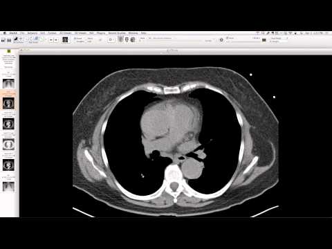
Chest radiographs show progression from diffuse, well-defined, upper-zone predominant 3-4 mm nodules (simple silicosis) to coalescence of nodules forming a large opacity, particularly in the left upper lobe, with associated volume loss in the upper lobes, upward hilar retraction, and emphysema in the lower lobes (complicated silicosis/progressive massive fibrosis). CT confirms perilymphatic distribution of pneumoconiotic nodules, pseudo-plaquing along pleural surfaces, and large, high-attenuation, calcified masses representing coalesced nodules. Hypertrophy of extrapleural fat and bilateral pleural plaques (consistent with asbestos exposure) are also noted.
Key takeaways
This case illustrates the progression from simple to complicated silicosis (progressive massive fibrosis) in a sandblaster and foundry worker. The classic findings include coalescing upper lobe nodules, volume loss, hilar retraction, and compensatory emphysema. Co-existing pleural plaques indicate additional asbestos exposure, common in foundry settings. Recognizing these patterns is crucial for occupational lung disease diagnosis.
AI-assisted summary — may contain errors. Verify against the source video. Learn more
More from this webinar
- Williams Syndrome with Supravalvular Aortic Stenosis and Pulmonary Artery NarrowingCongenital
- Alveolar Capillary Dysplasia with Unroofed Coronary Sinus (Right-to-Left Shunt)Congenital
- Iatrogenic Esophageal Rupture from Blakemore Tube PlacementIatrogenic
- Primum Atrial Septal Defect (ASD)Congenital
- Pulmonary Alveolar MicrolithiasisMetabolic
- Esophageal Varices Anastomosing with an Inferior Pulmonary VeinVascular
- Unroofed Coronary Sinus (Isolated Finding)Congenital
- Complex Congenital Anomalies: Right Aortic Arch with Aberrant Innominate Artery, Total Anomalous Pulmonary Venous Return of Left Lung, and Cor Triatriatum SinisterCongenital
See all cases from April 3, 2014 →
Related Exposure cases
- Centrilobular Emphysema Mimicking Lymphangioleiomyomatosis
- Chronic Beryllium Disease (Berylliosis) Mimicking Sarcoidosis
- Silicosis with Progressive Massive Fibrosis and Rounded Atelectasis
- Silicosis with Early Pseudoplaque and Eggshell Nodal Calcification
- Silicosis in a Hanford Site Construction Worker
- Accelerated countertop silicosis