 CTICases
Pulmonary Aspergilloma in Healed Post-Tuberculosis Cavity
 ▶ Watch on YouTube — age-restricted, cannot embed here
[...]. Pulmonary Aspergilloma in Healed Post-Tuberculosis Cavity. CTICases. Published April 3, 2014. https://cticases.com/c/8hjk7
Imaging findings
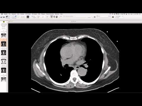
Chest radiograph shows progression from fibrocalcific scarring, volume loss, and bronchiectasis in the right upper lobe (consistent with healed tuberculosis) to the development of a new soft tissue nodule within this area. CT confirms a well-defined filling defect (aspergilloma) inside a thick-walled chronic cavity, surrounded by established bronchiectasis and calcification.
Key takeaways
This case illustrates a pulmonary aspergilloma developing within a pre-existing chronic cavity, a common sequela of healed tuberculosis, especially in regions with high TB prevalence. The aspergilloma causes local inflammation and can lead to symptoms like increased sputum production and cough. This serves as a reminder that worldwide, tuberculosis is the leading cause of chronic cavitary disease predisposing to aspergilloma formation.
AI-assisted summary — may contain errors. Verify against the source video. Learn more
More from this webinar
- Williams Syndrome with Supravalvular Aortic Stenosis and Pulmonary Artery NarrowingCongenital
- Alveolar Capillary Dysplasia with Unroofed Coronary Sinus (Right-to-Left Shunt)Congenital
- Iatrogenic Esophageal Rupture from Blakemore Tube PlacementIatrogenic
- Primum Atrial Septal Defect (ASD)Congenital
- Pulmonary Alveolar MicrolithiasisMetabolic
- Esophageal Varices Anastomosing with an Inferior Pulmonary VeinVascular
- Unroofed Coronary Sinus (Isolated Finding)Congenital
- Complex Congenital Anomalies: Right Aortic Arch with Aberrant Innominate Artery, Total Anomalous Pulmonary Venous Return of Left Lung, and Cor Triatriatum SinisterCongenital
See all cases from April 3, 2014 →