 CTICases
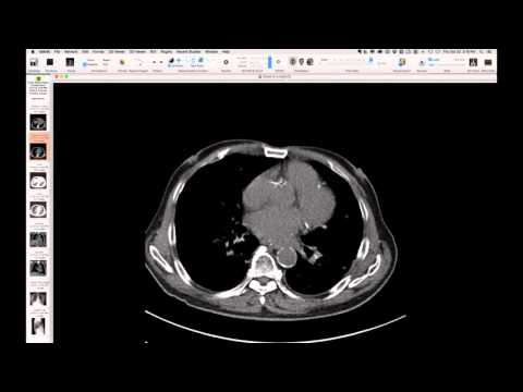
Defibrillator with CPR Efficacy Measurement Component
 ▶ Watch on YouTube — age-restricted, cannot embed here
[...]. Defibrillator with CPR Efficacy Measurement Component. CTICases. Published October 22, 2015. https://cticases.com/c/dkyhz
Imaging findings
Chest radiograph reveals a component of a defibrillator device not typically seen, appearing as a separate metallic structure integrated with the main apparatus. This component is designed to measure the efficacy and amplitude of chest compressions during CPR.
Key takeaways
This case highlights an unusual component of a modern defibrillator device (e.g., Zoll), which incorporates a feature to measure the efficacy of chest compressions during cardiopulmonary resuscitation. Recognizing this metallic component as part of an integrated defibrillation system, rather than a separate or misplaced foreign body, is important.
AI-assisted summary — may contain errors. Verify against the source video. Learn more
More from this webinar
- Interrupted Aortic Arch, Type A with Aortopulmonary WindowCongenital
- Interrupted Aortic Arch, Type B Variant with Aberrant Subclavian ArteriesCongenital
- Post-Transplant Lymphoproliferative Disorder (PTLD), Polyclonal Polymorphic TypeNeoplastic
- Primary Effusion Lymphoma (PFL) in HIV-Positive PatientNeoplastic
- Apical Hypertrophic Cardiomyopathy with Apical AneurysmCongenital
- Double Saddle Pulmonary EmbolismVascular
- Primary Coccidioidomycosis with CavitationInfection
- Disseminated CoccidioidomycosisInfection
See all cases from October 22, 2015 →