 CTICases
Apical Hypertrophic Cardiomyopathy with Apical Aneurysm
 ▶ Watch on YouTube — age-restricted, cannot embed here
[...]. Apical Hypertrophic Cardiomyopathy with Apical Aneurysm. CTICases. Published October 22, 2015. https://cticases.com/c/h3qpd
Imaging findings
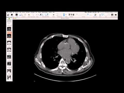
Cardiac imaging during diastole reveals normal heart morphology at the base, but towards the apex, there is a massive blob of markedly thick, circumferential myocardium with a hint of trabeculation, forming a large apical aneurysm. The coronary arteries are completely normal.
Key takeaways
This case demonstrates a massive apical aneurysm resulting from apical hypertrophic cardiomyopathy, emphasizing the distinctive imaging findings in contrast to an apical aneurysm caused by myocardial infarction. The presence of normal coronary arteries and marked apical myocardial hypertrophy are key features differentiating this variant of hypertrophic cardiomyopathy.
AI-assisted summary — may contain errors. Verify against the source video. Learn more
More from this webinar
- Interrupted Aortic Arch, Type A with Aortopulmonary WindowCongenital
- Interrupted Aortic Arch, Type B Variant with Aberrant Subclavian ArteriesCongenital
- Post-Transplant Lymphoproliferative Disorder (PTLD), Polyclonal Polymorphic TypeNeoplastic
- Primary Effusion Lymphoma (PFL) in HIV-Positive PatientNeoplastic
- Double Saddle Pulmonary EmbolismVascular
- Primary Coccidioidomycosis with CavitationInfection
- Disseminated CoccidioidomycosisInfection
- Isolated Pulmonary Cryptococcosis in an Immunocompetent PatientInfection
See all cases from October 22, 2015 →
Related Congenital cases
- Dual LAD Coronary Artery Anomaly
- Congenital Pulmonary Airway Malformation (CPAM) Mimicking Adenocarcinoma
- Bridging Bronchus with Azygos Fissure
- Hypertrophic Cardiomyopathy with Chordal Systolic Anterior Motion (SAM)
- Anomalous Left Main Coronary Artery with Intramural Course
- Congenitally Corrected Transposition of the Great Arteries (L-TGA)