 CTICases
Pulmonary Cavity with Aspergilloma and MAC Infection in COPD
 ▶ Watch on YouTube — age-restricted, cannot embed here
[...]. Pulmonary Cavity with Aspergilloma and MAC Infection in COPD. CTICases. Published April 3, 2014. https://cticases.com/c/u7smm
Imaging findings
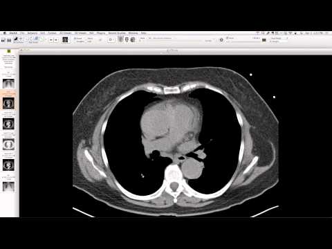
Chest radiograph reveals a soft tissue mass within a cavitary lesion in the right upper lobe. CT confirms a chronic cavity containing an aspergilloma, accompanied by severe emphysema. Hypertrophy of intercostal arteries and thickened extrapleural tissues are indicative of chronic inflammation. Mass-like areas of consolidation with calcification are also noted. Biopsy showed necrotizing granulomatous inflammation with positive AFB stain, confirmed as non-tuberculous mycobacteria (presumably MAC) via PCR.
Key takeaways
This case demonstrates the classic presentation of a pulmonary aspergilloma within a pre-existing chronic cavity, common in older men with COPD. The superimposition of a Mycobacterium avium complex (MAC) infection within areas of consolidation further complicates the picture. Differentiation from neoplasm is crucial, often requiring biopsy, which here revealed necrotizing granulomatous inflammation consistent with MAC.
AI-assisted summary — may contain errors. Verify against the source video. Learn more
More from this webinar
- Williams Syndrome with Supravalvular Aortic Stenosis and Pulmonary Artery NarrowingCongenital
- Alveolar Capillary Dysplasia with Unroofed Coronary Sinus (Right-to-Left Shunt)Congenital
- Iatrogenic Esophageal Rupture from Blakemore Tube PlacementIatrogenic
- Primum Atrial Septal Defect (ASD)Congenital
- Pulmonary Alveolar MicrolithiasisMetabolic
- Esophageal Varices Anastomosing with an Inferior Pulmonary VeinVascular
- Unroofed Coronary Sinus (Isolated Finding)Congenital
- Complex Congenital Anomalies: Right Aortic Arch with Aberrant Innominate Artery, Total Anomalous Pulmonary Venous Return of Left Lung, and Cor Triatriatum SinisterCongenital
See all cases from April 3, 2014 →