 CTICases
Acute Q Fever Pneumonia
 ▶ Watch on YouTube — age-restricted, cannot embed here
[...]. Acute Q Fever Pneumonia. CTICases. Published October 22, 2015. https://cticases.com/c/zcjxc
Imaging findings
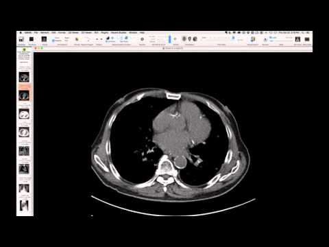
Initial chest radiograph showed hypoinflation. Subsequent radiographs revealed lower lobe consolidation and basal opacities, particularly after rapid progression to respiratory failure. CT imaging performed two weeks after presentation, despite motion artifact, suggested small nodules, dependent consolidation, ground glass opacities, and distinct subpleural lymphatic nodules, particularly along the fissures.
Key takeaways
This case illustrates acute Q fever (Coxiella burnetii pneumonia) in a dairy farm worker, characterized by acute respiratory failure, fever, and a notable bradycardia, which is a classic but often overlooked clinical sign. The imaging showed consolidating pneumonia and subtle lymphatic nodules. Q fever, a zoonosis transmitted by livestock, should be considered in the differential for severe community-acquired pneumonia unresponsive to standard antibiotics, especially in patients with relevant occupational exposures. It responds well to doxycycline.
AI-assisted summary — may contain errors. Verify against the source video. Learn more
More from this webinar
- Interrupted Aortic Arch, Type A with Aortopulmonary WindowCongenital
- Interrupted Aortic Arch, Type B Variant with Aberrant Subclavian ArteriesCongenital
- Post-Transplant Lymphoproliferative Disorder (PTLD), Polyclonal Polymorphic TypeNeoplastic
- Primary Effusion Lymphoma (PFL) in HIV-Positive PatientNeoplastic
- Apical Hypertrophic Cardiomyopathy with Apical AneurysmCongenital
- Double Saddle Pulmonary EmbolismVascular
- Primary Coccidioidomycosis with CavitationInfection
- Disseminated CoccidioidomycosisInfection
See all cases from October 22, 2015 →
Related Infection cases
- Disseminated Cryptococcosis in an Immunosuppressed Patient
- Cavitary Coccidioidomycosis Causing Pneumothorax
- Cervicofacial Actinomycosis with Mediastinal Extension
- Disseminated Nocardiosis with Cavitation in a Kidney Transplant Recipient
- Necrotizing Pneumonia (Group A Streptococcus) and Pneumocystis Pneumonia
- Coccidioidomycosis