 CTICases
Pseudo-Pulmonary Embolism (Mixing Phenomenon / Smoke Artifact)
 ▶ Watch on YouTube — age-restricted, cannot embed here
[...]. Pseudo-Pulmonary Embolism (Mixing Phenomenon / Smoke Artifact). CTICases. Published April 3, 2014. https://cticases.com/c/byqf2
Imaging findings
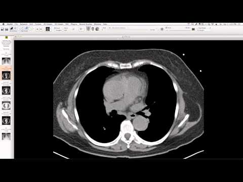
Extremely bright opacification (750-800+ HU) of the pulmonary arteries due to very early scanning and a leading bolus of contrast. Apparent intraluminal filling defects are noted, particularly conspicuous in the pulmonary arteries. Quantitative analysis of these "defects" reveals high Hounsfield units (e.g., 434-462 HU), indicating relatively less opacified blood mixing with highly opacified blood, rather than true low-attenuation thrombus.
Key takeaways
This case is a classic example of pseudo-pulmonary embolism, a common artifact caused by a mixing phenomenon of highly concentrated contrast with relatively unopacified blood in the pulmonary arteries, often seen with early-phase scanning. It's crucial to differentiate these "smoke artifacts" from true pulmonary emboli by assessing Hounsfield units within the apparent filling defects, as misinterpretation can lead to unnecessary treatment. The simultaneous brightness contrast illusion can also contribute to this visual perception.
AI-assisted summary — may contain errors. Verify against the source video. Learn more
More from this webinar
- Williams Syndrome with Supravalvular Aortic Stenosis and Pulmonary Artery NarrowingCongenital
- Alveolar Capillary Dysplasia with Unroofed Coronary Sinus (Right-to-Left Shunt)Congenital
- Iatrogenic Esophageal Rupture from Blakemore Tube PlacementIatrogenic
- Primum Atrial Septal Defect (ASD)Congenital
- Pulmonary Alveolar MicrolithiasisMetabolic
- Esophageal Varices Anastomosing with an Inferior Pulmonary VeinVascular
- Unroofed Coronary Sinus (Isolated Finding)Congenital
- Complex Congenital Anomalies: Right Aortic Arch with Aberrant Innominate Artery, Total Anomalous Pulmonary Venous Return of Left Lung, and Cor Triatriatum SinisterCongenital
See all cases from April 3, 2014 →
Related Artifact cases
- Pseudo-Nodule from Osteophyte with Reactive Scarring
- Pseudo-Pulmonary Embolism due to Bronchial Artery Flow Artifact
- AltaValve Transcatheter Mitral Valve Replacement
- Upside-down fluid level on CT from silicone breast implant rupture
- Anatomical Demonstration: Phrenic Nerve Course
- Pneumothorax Rupturing into Hernia Sac