 CTICases
Interstitial Lung Edema with Fluid in Interlobar Fissures
 ▶ Watch on YouTube — age-restricted, cannot embed here
[...]. Interstitial Lung Edema with Fluid in Interlobar Fissures. CTICases. Published October 22, 2015. https://cticases.com/c/s7vad
Imaging findings
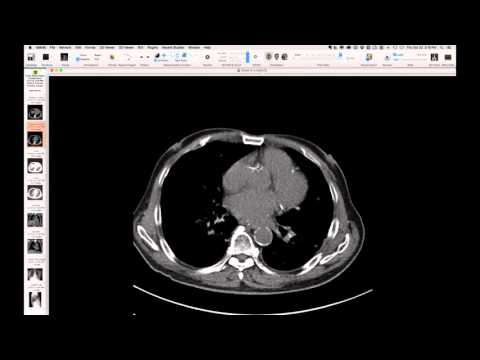
Frontal chest radiograph demonstrates conspicuous septal lines, including Kerley B lines in the bases and longer Kerley A lines. The lateral projection further reveals subpleural interstitial edema clearly related to the interlobar fissures, with actual fluid accumulation within the fissures themselves.
Key takeaways
This case provides an excellent illustration of interstitial lung edema, highlighting classic radiographic signs such as prominent septal (Kerley A and B) lines. Crucially, it demonstrates the progression from subpleural interstitial edema to the accumulation of free fluid directly within the interlobar fissures, a distinct manifestation of severe interstitial edema.
AI-assisted summary — may contain errors. Verify against the source video. Learn more
More from this webinar
- Interrupted Aortic Arch, Type A with Aortopulmonary WindowCongenital
- Interrupted Aortic Arch, Type B Variant with Aberrant Subclavian ArteriesCongenital
- Post-Transplant Lymphoproliferative Disorder (PTLD), Polyclonal Polymorphic TypeNeoplastic
- Primary Effusion Lymphoma (PFL) in HIV-Positive PatientNeoplastic
- Apical Hypertrophic Cardiomyopathy with Apical AneurysmCongenital
- Double Saddle Pulmonary EmbolismVascular
- Primary Coccidioidomycosis with CavitationInfection
- Disseminated CoccidioidomycosisInfection
See all cases from October 22, 2015 →
Related Vascular cases
- Chronic Pulmonary Thromboembolism (CTEPH)
- Reverse Takotsubo Cardiomyopathy
- Spontaneous Auto-Embolization of a Pulmonary Arteriovenous Malformation (AVM)
- Type A Aortic Dissection with Acute Bilateral Lower Extremity Paralysis and Retroperitoneal Hematoma
- Mycotic Aneurysm (from Enterococcus Faecalis Endocarditis)
- Left Ventricular Pseudoaneurysm