 CTICases
Squamous Cell Carcinoma of the Lung
 ▶ Watch on YouTube — age-restricted, cannot embed here
[...]. Squamous Cell Carcinoma of the Lung. CTICases. Published July 7, 2014. https://cticases.com/c/5rufx
Imaging findings
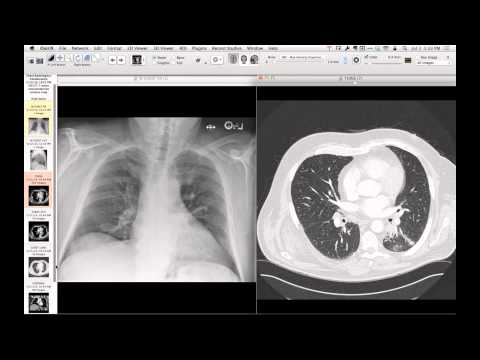
Chest CT shows a large, necrotic lung mass in the right lung. Dynamic contrast-enhanced magnetic resonance angiography demonstrates progressive, heterogeneous enhancement of the squamous cell carcinoma over successive phases, alongside an incidental filling defect in the right lower lobe pulmonary artery representing a pulmonary embolism.
Key takeaways
Dynamic contrast-enhanced MRI can be used to evaluate tumor perfusion and vascularity in lung cancers. MRA can also simultaneously evaluate the pulmonary vasculature for associated complications such as pulmonary embolism, which is common in hypercoagulable oncology patients.
AI-assisted summary — may contain errors. Verify against the source video. Learn more
More from this webinar
- Spontaneous Pneumomediastinum and Pneumoretroperitoneum due to Macklin PhenomenonOther
- Thoracic Spine Discitis-Osteomyelitis and Paraspinal Abscess due to Disseminated Mycobacterium avium complexInfection
- Right Ventricular Perforation and Pericardial Contrast InjectionIatrogenic
- Esophagopericardial FistulaOther
- Congenital Hypoplasia of the Right Upper LobeCongenital
- Goodpasture Syndrome (Anti-GBM Disease)Autoimmune
- Behçet Syndrome with Multiple Pulmonary Artery AneurysmsAutoimmune
- Left Ventricular Pseudoaneurysm post-TAVRIatrogenic
See all cases from July 7, 2014 →