 CTICases
Left Ventricular Pseudoaneurysm post-TAVR
 ▶ Watch on YouTube — age-restricted, cannot embed here
[...]. Left Ventricular Pseudoaneurysm post-TAVR. CTICases. Published July 7, 2014. https://cticases.com/c/6uk72
Imaging findings
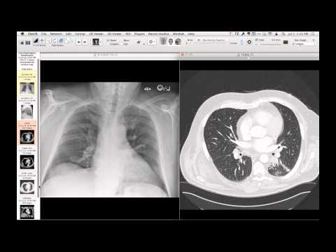
Contrast-enhanced chest CT demonstrates a lobulated pseudoaneurysm at the left ventricular apex with communication to the ventricular lumen, located near displaced surgical pledgets from a prior transapical TAVR site. Active contrast extravasation and accumulation are noted within the small apical aneurymsal sac.
Key takeaways
Apical pseudoaneurysm is a known mechanical complication of transapical access for transcatheter aortic valve replacement, often marked by the displacement or disruption of apical surgical closure pledgets. These pseudoaneurysms can be successfully treated with less invasive approaches, such as the percutaneous placement of an endovascular ventricular septal defect occluding device.
AI-assisted summary — may contain errors. Verify against the source video. Learn more
More from this webinar
- Spontaneous Pneumomediastinum and Pneumoretroperitoneum due to Macklin PhenomenonOther
- Thoracic Spine Discitis-Osteomyelitis and Paraspinal Abscess due to Disseminated Mycobacterium avium complexInfection
- Right Ventricular Perforation and Pericardial Contrast InjectionIatrogenic
- Esophagopericardial FistulaOther
- Congenital Hypoplasia of the Right Upper LobeCongenital
- Goodpasture Syndrome (Anti-GBM Disease)Autoimmune
- Behçet Syndrome with Multiple Pulmonary Artery AneurysmsAutoimmune
- Typical Bronchial CarcinoidNeoplastic
See all cases from July 7, 2014 →