 CTICases
Esophagopericardial Fistula
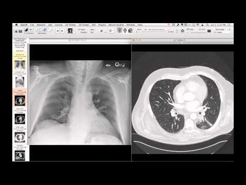 ▶ Watch on YouTube — age-restricted, cannot embed here
[...]. Esophagopericardial Fistula. CTICases. Published July 7, 2014. https://cticases.com/c/3cwme
Imaging findings
Chest radiography shows air in the mediastinum confined to the pericardial space, without extension superiorly into the neck recesses. An oral contrast pericardiogram, performed using barium or water-soluble contrast, demonstrates contrast extravasating from the lower esophagus directly into the pericardial space, confirming the presence of a fistula.
Key takeaways
Pneumopericardium can be differentiated from pneumomediastinum on radiography by the confinement of air below the level of the great vessel pericardial reflections. An esophagopericardial fistula is a rare, life-threatening complication that can develop after chemoradiation for esophageal cancer, and can be diagnosed elegantly using oral contrast administration to document communication between the GI tract and the pericardium.
AI-assisted summary — may contain errors. Verify against the source video. Learn more
More from this webinar
- Spontaneous Pneumomediastinum and Pneumoretroperitoneum due to Macklin PhenomenonOther
- Thoracic Spine Discitis-Osteomyelitis and Paraspinal Abscess due to Disseminated Mycobacterium avium complexInfection
- Right Ventricular Perforation and Pericardial Contrast InjectionIatrogenic
- Congenital Hypoplasia of the Right Upper LobeCongenital
- Goodpasture Syndrome (Anti-GBM Disease)Autoimmune
- Behçet Syndrome with Multiple Pulmonary Artery AneurysmsAutoimmune
- Left Ventricular Pseudoaneurysm post-TAVRIatrogenic
- Typical Bronchial CarcinoidNeoplastic
See all cases from July 7, 2014 →