 CTICases
Right Ventricular Perforation and Pericardial Contrast Injection
 ▶ Watch on YouTube — age-restricted, cannot embed here
[...]. Right Ventricular Perforation and Pericardial Contrast Injection. CTICases. Published July 7, 2014. https://cticases.com/c/w2r8s
Imaging findings
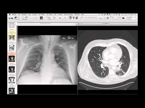
Chest radiography shows a newly placed subclavian central venous line positioned deep within the right atrium or right ventricle. CT imaging demonstrates a large volume of contrast medium filling the pericardial space, beautifully outlining the pericardial anatomy including the transverse sinus, superior aortic recesses, pulmonary recesses, and the posterior oblique sinus, rather than distributing normally through the systemic vasculature.
Key takeaways
Central venous catheters positioned deep within the heart can abut the myocardial wall and cause transient perforation during pressurized injection of contrast, leading to iatrogenic intrapericardial contrast administration. Acutely, the instillation of even a small volume of fluid within the non-distensible fibrous pericardium can cause cardiac tamponade, though patients can occasionally remain hemodynamically stable and require only close observation.
AI-assisted summary — may contain errors. Verify against the source video. Learn more
More from this webinar
- Spontaneous Pneumomediastinum and Pneumoretroperitoneum due to Macklin PhenomenonOther
- Thoracic Spine Discitis-Osteomyelitis and Paraspinal Abscess due to Disseminated Mycobacterium avium complexInfection
- Esophagopericardial FistulaOther
- Congenital Hypoplasia of the Right Upper LobeCongenital
- Goodpasture Syndrome (Anti-GBM Disease)Autoimmune
- Behçet Syndrome with Multiple Pulmonary Artery AneurysmsAutoimmune
- Left Ventricular Pseudoaneurysm post-TAVRIatrogenic
- Typical Bronchial CarcinoidNeoplastic
See all cases from July 7, 2014 →