 CTICases
Chest Wall Hematoma with Active Extravasation
 ▶ Watch on YouTube — age-restricted, cannot embed here
[...]. Chest Wall Hematoma with Active Extravasation. CTICases. Published July 17, 2014. https://cticases.com/c/6mfct
Imaging findings
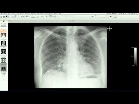
Chest radiograph demonstrates asymmetric density across the entire left hemithorax. Contrast-enhanced chest CT reveals an extremely large hematoma in the left lateral and posterior chest wall with foci of active arterial extravasation, a small hemothorax, and dependent atelectasis. Angiography confirms active bleeding from intercostal and chest wall arteries, which were successfully embolized.
Key takeaways
Massive chest wall hematomas can occur after minor trauma in patients who are super-therapeutic on anticoagulants (such as warfarin, with an INR of 9.9 in this case). On a chest radiograph, this can present as a diffuse asymmetric hemithorax opacity that mimics a large pleural effusion or parenchymal process; CT is essential to identify the chest wall origin and any active arterial extravasation amenable to transcatheter embolization.
AI-assisted summary — may contain errors. Verify against the source video. Learn more
More from this webinar
- Aortic Arch DissectionVascular
- Sclerosing PeriaortitisInflammatory
- Hypoplastic Left Heart Syndrome (Post-Norwood and Sano Shunt)Congenital
- Tetralogy of Fallot with Ductal Origin of the Left Pulmonary ArteryCongenital
- Migrating Pleural Mesothelial CystCongenital
- Chronic Diaphragmatic RuptureTrauma
- Bronchopulmonary DysplasiaCongenital
- Granulomatous-Lymphocytic Interstitial Lung DiseaseILD
See all cases from July 17, 2014 →