 CTICases
Sclerosing Periaortitis
 ▶ Watch on YouTube — age-restricted, cannot embed here
[...]. Sclerosing Periaortitis. CTICases. Published July 17, 2014. https://cticases.com/c/hynev
Imaging findings
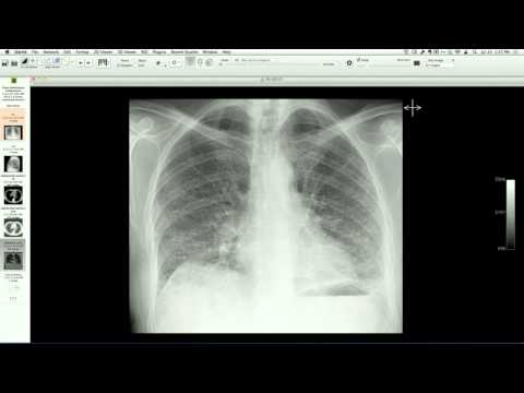
CT angiography demonstrates circumferential soft-tissue thickening around the ascending aorta extending into the arch, with abnormal soft tissue surrounding the left subclavian artery. Non-contrast CT reveals high-density material and calcification within the outer layers of the aortic wall (adventitia and media) without inward displacement of the calcification, distinguishing it from an intramural hematoma.
Key takeaways
Sclerosing periaortitis is an inflammatory condition of the aortic wall that can clinically and radiologically mimic an intramural hematoma or typical large-vessel vasculitis like Giant Cell or Takayasu arteritis. Pathologically distinct, it primarily involves adventitial and medial inflammation rather than intimal displacement, and is characterized by dense fibrosis and inflammatory infiltration.
AI-assisted summary — may contain errors. Verify against the source video. Learn more
More from this webinar
- Aortic Arch DissectionVascular
- Hypoplastic Left Heart Syndrome (Post-Norwood and Sano Shunt)Congenital
- Tetralogy of Fallot with Ductal Origin of the Left Pulmonary ArteryCongenital
- Migrating Pleural Mesothelial CystCongenital
- Chronic Diaphragmatic RuptureTrauma
- Bronchopulmonary DysplasiaCongenital
- Granulomatous-Lymphocytic Interstitial Lung DiseaseILD
- Aortic Coarctation with Bicuspid Aortic ValveCongenital
See all cases from July 17, 2014 →