 CTICases
Migrating Pleural Mesothelial Cyst
 ▶ Watch on YouTube — age-restricted, cannot embed here
[...]. Migrating Pleural Mesothelial Cyst. CTICases. Published July 17, 2014. https://cticases.com/c/5a6f7
Imaging findings
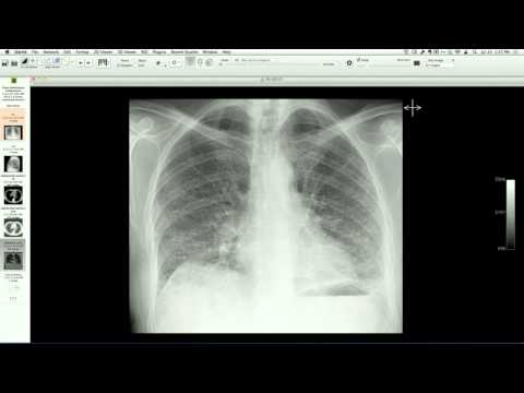
Serial imaging including chest CT, MRI, and PET/CT demonstrates a benign, fluid-filled cystic structure in the pleural space that migrates between different locations on sequential examinations, including the anterior mediastinum, the oblique fissure, and the posterior pleural space. Surgical exploration confirms a benign mesothelial cyst attached to the pleural surface by a very thin, pedunculated stock.
Key takeaways
Mesothelial cysts of the pleural space are benign lesions that can occasionally be pedunculated, allowing them to freely migrate within the pleural cavity or fissures. This mobility can lead to diagnostic confusion, prompting unnecessary biopsies or concerns for malignancy if the cyst's changing position on serial scans is not recognized as a hallmark of a "wandering" or migrating pleural cyst.
AI-assisted summary — may contain errors. Verify against the source video. Learn more
More from this webinar
- Aortic Arch DissectionVascular
- Sclerosing PeriaortitisInflammatory
- Hypoplastic Left Heart Syndrome (Post-Norwood and Sano Shunt)Congenital
- Tetralogy of Fallot with Ductal Origin of the Left Pulmonary ArteryCongenital
- Chronic Diaphragmatic RuptureTrauma
- Bronchopulmonary DysplasiaCongenital
- Granulomatous-Lymphocytic Interstitial Lung DiseaseILD
- Aortic Coarctation with Bicuspid Aortic ValveCongenital
See all cases from July 17, 2014 →