 CTICases
Chronic Diaphragmatic Rupture
 ▶ Watch on YouTube — age-restricted, cannot embed here
[...]. Chronic Diaphragmatic Rupture. CTICases. Published July 17, 2014. https://cticases.com/c/ws7u4
Imaging findings
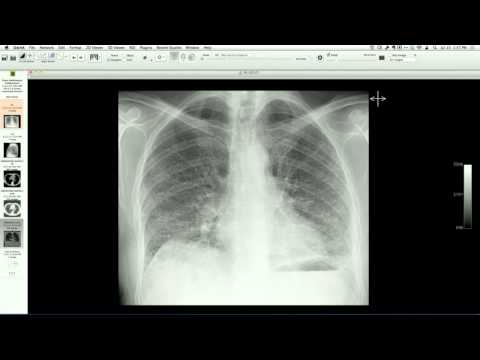
Chest CT demonstrates a defect in the left hemidiaphragm with herniation of intra-abdominal fat and colon into the left hemithorax. The "dependent viscera sign" is present, where herniated abdominal organs lie directly against the posterior ribs due to the loss of diaphragmatic support. Multiple healed left-sided rib fractures are also noted.
Key takeaways
Diaphragmatic rupture can occur after blunt trauma and may go undetected acutely, presenting years later as a chronic diaphragmatic hernia. Because of the negative intrathoracic pressure, abdominal contents can progressively herniate into the thorax over time, potentially causing respiratory distress or bowel obstruction, and necessitating surgical repair with mesh or primary closure.
AI-assisted summary — may contain errors. Verify against the source video. Learn more
More from this webinar
- Aortic Arch DissectionVascular
- Sclerosing PeriaortitisInflammatory
- Hypoplastic Left Heart Syndrome (Post-Norwood and Sano Shunt)Congenital
- Tetralogy of Fallot with Ductal Origin of the Left Pulmonary ArteryCongenital
- Migrating Pleural Mesothelial CystCongenital
- Bronchopulmonary DysplasiaCongenital
- Granulomatous-Lymphocytic Interstitial Lung DiseaseILD
- Aortic Coarctation with Bicuspid Aortic ValveCongenital
See all cases from July 17, 2014 →