 CTICases
Typical Bronchial Carcinoid
 ▶ Watch on YouTube — age-restricted, cannot embed here
[...]. Typical Bronchial Carcinoid. CTICases. Published July 7, 2014. https://cticases.com/c/98sra
Imaging findings
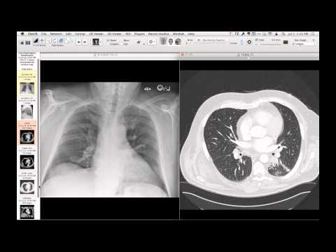
Outpatient chest radiography shows a subtle, irregularly shaped opacity beneath the left main bronchus. Chest CT confirms a rounded, vascular, endobronchial mass in the superior segment of the left lower lobe, causing adjacent lung atelectasis. Bronchoscopy correlates with a highly vascular, smooth lobulated mass that nearly completely occludes the bronchial lumen.
Key takeaways
Bronchial carcinoids are slow-growing, highly vascular neuroendocrine tumors that often present as endobronchial masses. They frequently present with recurrent post-obstructive pneumonia or hemoptysis, but can be discovered incidentally on radiographs as subtle subcarinal or retrocardiac opacities.
AI-assisted summary — may contain errors. Verify against the source video. Learn more
More from this webinar
- Spontaneous Pneumomediastinum and Pneumoretroperitoneum due to Macklin PhenomenonOther
- Thoracic Spine Discitis-Osteomyelitis and Paraspinal Abscess due to Disseminated Mycobacterium avium complexInfection
- Right Ventricular Perforation and Pericardial Contrast InjectionIatrogenic
- Esophagopericardial FistulaOther
- Congenital Hypoplasia of the Right Upper LobeCongenital
- Goodpasture Syndrome (Anti-GBM Disease)Autoimmune
- Behçet Syndrome with Multiple Pulmonary Artery AneurysmsAutoimmune
- Left Ventricular Pseudoaneurysm post-TAVRIatrogenic
See all cases from July 7, 2014 →