 CTICases
Focal Pulmonary Fibrosis
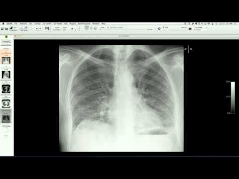 ▶ Watch on YouTube — age-restricted, cannot embed here
[...]. Focal Pulmonary Fibrosis. CTICases. Published July 17, 2014. https://cticases.com/c/8gsn5
Imaging findings
CT angiography demonstrates a persistent ground-glass lesion in the middle lobe with cystic components and bubble-like lucencies, along with a separate nodular ground-glass lesion in the lower lobe. Wedge resection of the middle lobe reveals focal chronic lymphocytic infiltration with fibrosis and no evidence of adenocarcinoma.
Key takeaways
Focal pulmonary fibrosis and localized chronic lymphocytic inflammation can present on chest CT as persistent ground-glass lesions containing cystic or bubble-like lucencies, highly mimicking low-grade adenocarcinoma (such as minimally invasive adenocarcinoma or adenocarcinoma in situ). Histological examination is often required for definitive diagnosis when these lesions persist or grow.
AI-assisted summary — may contain errors. Verify against the source video. Learn more
More from this webinar
- Aortic Arch DissectionVascular
- Sclerosing PeriaortitisInflammatory
- Hypoplastic Left Heart Syndrome (Post-Norwood and Sano Shunt)Congenital
- Tetralogy of Fallot with Ductal Origin of the Left Pulmonary ArteryCongenital
- Migrating Pleural Mesothelial CystCongenital
- Chronic Diaphragmatic RuptureTrauma
- Bronchopulmonary DysplasiaCongenital
- Granulomatous-Lymphocytic Interstitial Lung DiseaseILD
See all cases from July 17, 2014 →