 CTICases
Nocardiosis in a Heart Transplant Patient
 ▶ Watch on YouTube — age-restricted, cannot embed here
[...]. Nocardiosis in a Heart Transplant Patient. CTICases. Published March 4, 2016. https://cticases.com/c/9ca45
Imaging findings
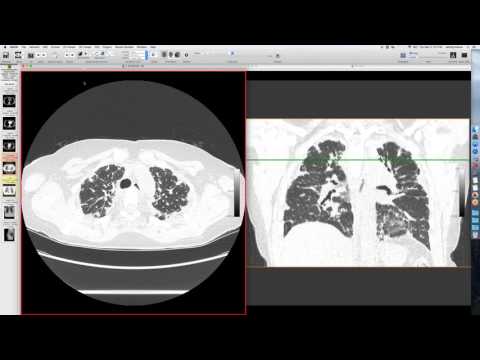
An initial radiograph was available. A CT performed for pulmonary embolism also showed a peripheral area of consolidation and ground glass on the right, as well as a cavitary lesion in the lower lobe with a relatively thin wall and some debris. A later CT revealed new nodularity with cavitation and satellite nodules, appearing granulomatous and airway-centered. The cavity wall became thinner over time.
Key takeaways
In immune-suppressed patients, Nocardia tends to present as larger lesions and often manages more like a fungal infection, mimicking cryptococcus or coccidioidomycosis. The patient was treated for community-acquired pneumonia but did not respond, with nocardiosis later confirmed. Nocardia responds to sulfa drugs, and its incidence has decreased due to widespread Pneumocystis prophylaxis. Cases are typically seen in transplant patients or those with lupus.
AI-assisted summary — may contain errors. Verify against the source video. Learn more
More from this webinar
- Fat Embolism SyndromeTrauma
- Dystrophic Calcification from Rhabdomyolysis and Pulmonary Interstitial EmphysemaMetabolic
- Pleuroparenchymal Fibroelastosis as a Manifestation of Graft-versus-Host DiseaseILD
- Synchronous Lung Cancer and Pulmonary HamartomaNeoplastic
- Variant Coronary Artery in the Ligament of MarshallCongenital
- Job's Syndrome (Hyper-IgE Syndrome) with Chronic Staphylococcal InfectionsAutoimmune
- Squamous Cell Carcinoma Obstructing Bronchus IntermediusNeoplastic
- Metastatic Breast Cancer Mimicking Primary Lung CancerNeoplastic
See all cases from March 4, 2016 →
Related Infection cases
- Coccidioidomycosis Presenting as Lobar Pneumonia in a Renal Transplant Patient
- Achalasia Complicated by Mycobacterium fortuitum Pneumonia
- Coxsackie Viral Constrictive Pericarditis
- Pulmonary Coccidioidomycosis Mimicking Metastasis
- Recurrent Infective Endocarditis with Mycotic Pulmonary Aneurysms
- Discitis-Osteomyelitis