 CTICases
Squamous Cell Carcinoma Obstructing Bronchus Intermedius
 ▶ Watch on YouTube — age-restricted, cannot embed here
[...]. Squamous Cell Carcinoma Obstructing Bronchus Intermedius. CTICases. Published March 4, 2016. https://cticases.com/c/d6g6g
Imaging findings
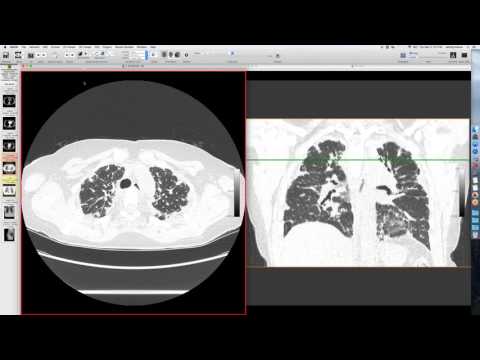
A 50-year-old male presented for lung cancer screening. A coronal thick-section mean image (pseudo chest X-ray) showed volume loss on the right, with clear obstruction of the bronchus intermedius. Further CT confirmed an endobronchial lesion.
Key takeaways
This was the most dramatic lung cancer seen on a screening CT, clearly indicating an obstructing endobronchial lesion. It was categorized as a Lung-RADS 4B with an X modifier, with near-certainty of malignancy. PET confirmed a hypermetabolic lesion, which turned out to be squamous cell carcinoma. Despite the extensive findings, the patient's symptoms were minimal, with only slight hemoptysis and shortness of breath.
AI-assisted summary — may contain errors. Verify against the source video. Learn more
More from this webinar
- Fat Embolism SyndromeTrauma
- Dystrophic Calcification from Rhabdomyolysis and Pulmonary Interstitial EmphysemaMetabolic
- Nocardiosis in a Heart Transplant PatientInfection
- Pleuroparenchymal Fibroelastosis as a Manifestation of Graft-versus-Host DiseaseILD
- Synchronous Lung Cancer and Pulmonary HamartomaNeoplastic
- Variant Coronary Artery in the Ligament of MarshallCongenital
- Job's Syndrome (Hyper-IgE Syndrome) with Chronic Staphylococcal InfectionsAutoimmune
- Metastatic Breast Cancer Mimicking Primary Lung CancerNeoplastic
See all cases from March 4, 2016 →
Related Neoplastic cases
- Diffuse Large B-cell Lymphoma with Myocardial and Pulmonary Involvement
- Thymoma with Paraneoplastic Pure Red Cell Aplasia
- Hidden Lung Cancer
- Pulmonary Adenocarcinoma with Retained Acupuncture Needles
- Extensive Lymphoma with Extrapleural Paraspinal Involvement
- Obstructive Left Upper Lobe Collapse (Carcinoid and Adenocarcinoma)