 CTICases
Fat Embolism Syndrome
 ▶ Watch on YouTube — age-restricted, cannot embed here
[...]. Fat Embolism Syndrome. CTICases. Published March 4, 2016. https://cticases.com/c/pdk68
Imaging findings
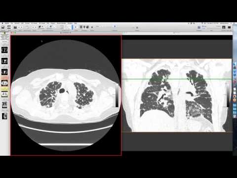
His initial radiograph showed good appearance, but he had a bad femoral fracture and a tibial fracture. After orthopedic surgery, he became short of breath. A CT angio showed pulmonary embolism with clot, and extensive consolidation and ground-glass opacity in the lungs, some consolidative and centered on the lobule, and more confluent in other areas.
Key takeaways
In this setting, the presumptive diagnosis was fat embolism with PE. The opacities cleared in about 2 days with supportive therapy, and he was anticoagulated for PE. This is more common in younger males with lower extremity long bone fractures or pelvic fractures, often presenting after surgery, possibly related to increased pressure or macroscopic fat embolization during nail placement. While literature cites high mortality, it's typically well-managed with supportive therapy now. Patients often experience hypoxia and may also develop petechial rashes and altered mental status.
AI-assisted summary — may contain errors. Verify against the source video. Learn more
More from this webinar
- Dystrophic Calcification from Rhabdomyolysis and Pulmonary Interstitial EmphysemaMetabolic
- Nocardiosis in a Heart Transplant PatientInfection
- Pleuroparenchymal Fibroelastosis as a Manifestation of Graft-versus-Host DiseaseILD
- Synchronous Lung Cancer and Pulmonary HamartomaNeoplastic
- Variant Coronary Artery in the Ligament of MarshallCongenital
- Job's Syndrome (Hyper-IgE Syndrome) with Chronic Staphylococcal InfectionsAutoimmune
- Squamous Cell Carcinoma Obstructing Bronchus IntermediusNeoplastic
- Metastatic Breast Cancer Mimicking Primary Lung CancerNeoplastic
See all cases from March 4, 2016 →