 CTICases
Cardiogenic Pulmonary Edema with Ground Glass Opacity
 ▶ Watch on YouTube — age-restricted, cannot embed here
[...]. Cardiogenic Pulmonary Edema with Ground Glass Opacity. CTICases. Published August 20, 2015. https://cticases.com/c/9jmcz
Imaging findings
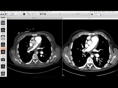
Radiograph showed a few septal lines. CT showed beautiful septal lines and areas of lobular ground glass opacity in the periphery of the lung. Effusions were also present. Occasional candidates for "lakes" (central lobular artery with engorged vein/lymphatic) were observed.
Key takeaways
Cardiogenic pulmonary edema can present with classic septal lines and, less commonly, beautiful lobular ground glass opacities in the lung periphery. These ground glass opacities can be seen along veins and lymphatics, sometimes presenting as "lakes." This is a characteristic appearance for teaching purposes.
AI-assisted summary — may contain errors. Verify against the source video. Learn more
More from this webinar
- Retained Wires from First-Generation DefibrillatorIatrogenic
- Hyperacute Pulmonary Infarction progressing to Acute PE with Pleural EffusionVascular
- Left Upper Lobe Anomalous Pulmonary Venous Stricture (Incidental)Congenital
- Lymphocytic Interstitial Pneumonia (LIP) in association with B-cell LymphomaNeoplastic
- Endobronchial Papillomatosis with Left Lower Lobe CollapseNeoplastic
- Esophageal Carcinoma with Radiographic CorrelationNeoplastic
- Hypersensitivity Pneumonitis (HP) in a Dog GroomerILD
- Auto-pneumonectomy and SVC Obstruction due to Necrotizing InfectionVascular
See all cases from August 20, 2015 →
Related Vascular cases
- Complex Arteriovenous Malformation (Internal Mammary Artery Supply)
- Chronic Thromboembolic Pulmonary Hypertension (CTEPH) - Peripheral Disease
- Retrograde Extension of Type B Aortic Dissection into the Aortic Arch with Endovascular Repair
- Ruptured Type B Aortic Intramural Hematoma with Hemothorax
- Systolic Anterior Motion (SAM) of the Mitral Valve without Hypertrophic Cardiomyopathy
- Cavitating Pulmonary Infarct secondary to Pulmonary Embolism