 CTICases
Esophageal Carcinoma with Radiographic Correlation
 ▶ Watch on YouTube — age-restricted, cannot embed here
[...]. Esophageal Carcinoma with Radiographic Correlation. CTICases. Published August 20, 2015. https://cticases.com/c/7b2kw
Imaging findings
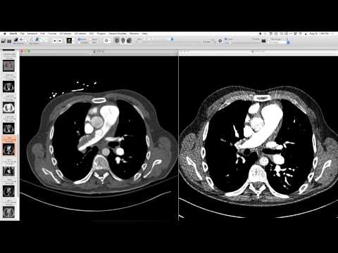
Frontal radiograph showed an extra bump in the lower mediastinum, with the descending aorta and azygos esophageal recess appearing convex. Lateral view was non-contributory. CT correlation revealed fluid and dilation in the esophagus, and a clearly abnormal, thickened esophagus with a narrow lumen protruding to the left, and a large lesion. Esophagram confirmed irregular shape, classic for carcinoma.
Key takeaways
Esophageal carcinoma can manifest on chest radiographs as subtle mediastinal contour abnormalities, such as an extra bump in the lower mediastinum or convexity of the azygos esophageal recess. CT and esophagram provide definitive diagnosis by showing esophageal thickening, luminal narrowing, and tumor mass.
AI-assisted summary — may contain errors. Verify against the source video. Learn more
More from this webinar
- Retained Wires from First-Generation DefibrillatorIatrogenic
- Hyperacute Pulmonary Infarction progressing to Acute PE with Pleural EffusionVascular
- Left Upper Lobe Anomalous Pulmonary Venous Stricture (Incidental)Congenital
- Lymphocytic Interstitial Pneumonia (LIP) in association with B-cell LymphomaNeoplastic
- Endobronchial Papillomatosis with Left Lower Lobe CollapseNeoplastic
- Cardiogenic Pulmonary Edema with Ground Glass OpacityVascular
- Hypersensitivity Pneumonitis (HP) in a Dog GroomerILD
- Auto-pneumonectomy and SVC Obstruction due to Necrotizing InfectionVascular
See all cases from August 20, 2015 →
Related Neoplastic cases
- Choriocarcinoma with Hemorrhagic Pulmonary Metastases
- Adenocarcinoma Spectrum (Preinvasive and Invasive Components)
- Pericardial Vascular Malformation (Veno-Hemangioma with Phleboliths)
- Recurrent Squamous Cell Carcinoma in Respiratory Papillomatosis with Rib Invasion
- Ectopic Mediastinal Parathyroid Adenoma
- Post-Transplant Lymphoproliferative Disorder (PTLD) Presenting as Airway-Centric Lung Nodules