 CTICases
Lymphocytic Interstitial Pneumonia (LIP) in association with B-cell Lymphoma
 ▶ Watch on YouTube — age-restricted, cannot embed here
[...]. Lymphocytic Interstitial Pneumonia (LIP) in association with B-cell Lymphoma. CTICases. Published August 20, 2015. https://cticases.com/c/qt4rn
Imaging findings
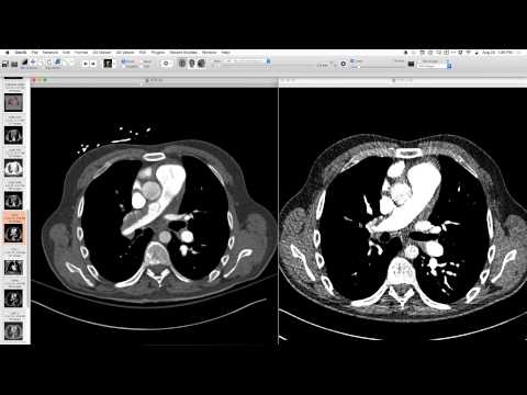
Initial CT showed extensive, bulky, partially confluent mediastinal, hilar, subcarinal, and intralobar lymphadenopathy associated with B-cell lymphoma. Extensive opacification throughout the lungs with multiple thin-walled cystic lesions in a perivascular distribution, increasing in number and size towards the lung bases, consistent with LIP. Follow-up CT after treatment showed improving opacification, residual nodularity, and persistent cystic lesions with characteristic appearance. Abdominal windows showed improved lymphadenopathy post-treatment.
Key takeaways
Lymphocytic Interstitial Pneumonia (LIP) can be associated with B-cell lymphoma. The imaging findings include extensive pulmonary opacification and thin-walled cystic lesions, increasing in size and number from upper to lower lungs. Mediastinal lymphadenopathy in these cases can be bulky and lymphoma-like. Cystic spaces are common in B-cell tumors due to protein production, and while often associated with LIP, they can occur independently.
AI-assisted summary — may contain errors. Verify against the source video. Learn more
More from this webinar
- Retained Wires from First-Generation DefibrillatorIatrogenic
- Hyperacute Pulmonary Infarction progressing to Acute PE with Pleural EffusionVascular
- Left Upper Lobe Anomalous Pulmonary Venous Stricture (Incidental)Congenital
- Endobronchial Papillomatosis with Left Lower Lobe CollapseNeoplastic
- Esophageal Carcinoma with Radiographic CorrelationNeoplastic
- Cardiogenic Pulmonary Edema with Ground Glass OpacityVascular
- Hypersensitivity Pneumonitis (HP) in a Dog GroomerILD
- Auto-pneumonectomy and SVC Obstruction due to Necrotizing InfectionVascular
See all cases from August 20, 2015 →
Related Neoplastic cases
- Choriocarcinoma with Hemorrhagic Pulmonary Metastases
- Adenocarcinoma Spectrum (Preinvasive and Invasive Components)
- Pericardial Vascular Malformation (Veno-Hemangioma with Phleboliths)
- Recurrent Squamous Cell Carcinoma in Respiratory Papillomatosis with Rib Invasion
- Ectopic Mediastinal Parathyroid Adenoma
- Post-Transplant Lymphoproliferative Disorder (PTLD) Presenting as Airway-Centric Lung Nodules