 CTICases
Hypersensitivity Pneumonitis (HP) in a Dog Groomer
 ▶ Watch on YouTube — age-restricted, cannot embed here
[...]. Hypersensitivity Pneumonitis (HP) in a Dog Groomer. CTICases. Published August 20, 2015. https://cticases.com/c/bwhza
Imaging findings
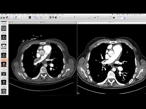
Initial radiograph showed too-dense lungs, ill-defined vessels, and questioned underlying ground glass opacity. Follow-up CT a week and a half later revealed diffuse ground glass opacity, very central lobular, with some lobules appearing very black, indicating mosaic attenuation and presumably air trapping. A small cyst was also noted.
Key takeaways
Hypersensitivity pneumonitis (HP) is characterized by diffuse ground glass opacity, often central lobular, with mosaic attenuation and air trapping. Radiographs can show subtle increased lung density and ill-defined vasculature. Identifying the antigen is crucial for prognosis, as continued exposure worsens outcomes. Mammalian antigens are rare causes; avian or amphibian exposures are more common, or even water sources with MAC.
AI-assisted summary — may contain errors. Verify against the source video. Learn more
More from this webinar
- Retained Wires from First-Generation DefibrillatorIatrogenic
- Hyperacute Pulmonary Infarction progressing to Acute PE with Pleural EffusionVascular
- Left Upper Lobe Anomalous Pulmonary Venous Stricture (Incidental)Congenital
- Lymphocytic Interstitial Pneumonia (LIP) in association with B-cell LymphomaNeoplastic
- Endobronchial Papillomatosis with Left Lower Lobe CollapseNeoplastic
- Esophageal Carcinoma with Radiographic CorrelationNeoplastic
- Cardiogenic Pulmonary Edema with Ground Glass OpacityVascular
- Auto-pneumonectomy and SVC Obstruction due to Necrotizing InfectionVascular
See all cases from August 20, 2015 →
Related ILD cases
- Methotrexate-induced Lung Toxicity (Organizing Pneumonia)
- Systemic Sclerosis-associated Nonspecific Interstitial Pneumonia (NSIP) - Progression
- Desquamative Interstitial Pneumonia (DIP)
- Amiodarone-induced Organizing Pneumonia
- Fibrotic Hypersensitivity Pneumonitis
- Alveolar Proteinosis with Complete Response to GM-CSF Therapy