 CTICases
Retained Wires from First-Generation Defibrillator
 ▶ Watch on YouTube — age-restricted, cannot embed here
[...]. Retained Wires from First-Generation Defibrillator. CTICases. Published August 20, 2015. https://cticases.com/c/u38b6
Imaging findings
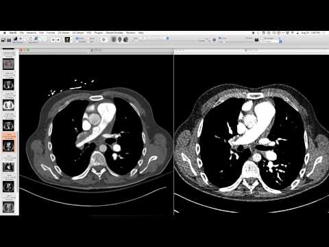
Frontal chest x-ray showed sternotomy wires in the shape of a square, with epicardial leads. Lateral projection showed wires anterior and posterior to the heart. These were identified as rectangular wire patches containing a very fine mesh of lines that do not resolve on conventional radiographs, along with a dense bar on the lead.
Key takeaways
These are retained wires from first-generation implanted defibrillators from the 1980s, which required open surgery and direct implantation. The pulse generators were large and implanted in the lower abdomen or pelvis. Later generations used oval patches with coarser wires. These are also known as patch electrodes.
AI-assisted summary — may contain errors. Verify against the source video. Learn more
More from this webinar
- Hyperacute Pulmonary Infarction progressing to Acute PE with Pleural EffusionVascular
- Left Upper Lobe Anomalous Pulmonary Venous Stricture (Incidental)Congenital
- Lymphocytic Interstitial Pneumonia (LIP) in association with B-cell LymphomaNeoplastic
- Endobronchial Papillomatosis with Left Lower Lobe CollapseNeoplastic
- Esophageal Carcinoma with Radiographic CorrelationNeoplastic
- Cardiogenic Pulmonary Edema with Ground Glass OpacityVascular
- Hypersensitivity Pneumonitis (HP) in a Dog GroomerILD
- Auto-pneumonectomy and SVC Obstruction due to Necrotizing InfectionVascular
See all cases from August 20, 2015 →
Related Iatrogenic cases
- Arterial Central Venous Catheter Placement (into aorta) via IJ and Subclavian Vein
- Retained Suction Catheter Tip (Intraoperative Foreign Body)
- Bilateral Capnothorax Post Heller Myotomy and Nissen Fundoplication
- Myocardial Perforation by Pacemaker Lead with Insulation Disruption
- Iatrogenic NG Tube Malposition with Mediastinal and Pleural Perforation
- Valve-in-Valve TAVR for Paravalvular Leak