 CTICases
Pulmonary Langerhans Cell Histiocytosis with Superimposed Haemophilus influenzae Bronchiolitis
 ▶ Watch on YouTube — age-restricted, cannot embed here
[...]. Pulmonary Langerhans Cell Histiocytosis with Superimposed Haemophilus influenzae Bronchiolitis. CTICases. Published April 3, 2014. https://cticases.com/c/hpa5w
Imaging findings
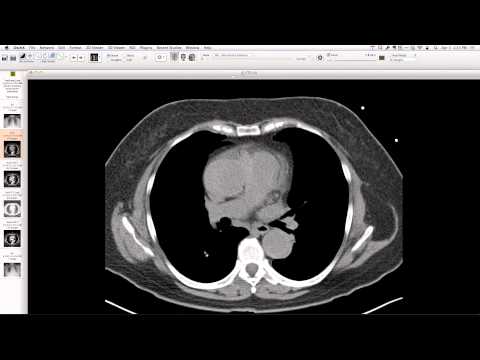
Chest radiograph shows ill-defined, smudgy, fuzzy nodules predominantly in the lower lungs with indistinct vessels. CT reveals extensive tree-in-bud opacities consistent with bronchiolitis, along with numerous thin-walled cysts and some emphysema, primarily in the upper and mid-lungs, sparing the bases.
Key takeaways
This case highlights the co-existence of Pulmonary Langerhans Cell Histiocytosis (PLCH), strongly associated with smoking, and a superimposed acute infection (Haemophilus influenzae bronchiolitis). PLCH typically presents with characteristic cysts and nodules, predominantly in the upper lobes, which can be distinguished from the infectious tree-in-bud pattern. While acute infection requires treatment, cessation of smoking is key for managing PLCH, with follow-up imaging to assess resolution of the infectious component and stability of the underlying cystic changes.
AI-assisted summary — may contain errors. Verify against the source video. Learn more
More from this webinar
- Williams Syndrome with Supravalvular Aortic Stenosis and Pulmonary Artery NarrowingCongenital
- Alveolar Capillary Dysplasia with Unroofed Coronary Sinus (Right-to-Left Shunt)Congenital
- Iatrogenic Esophageal Rupture from Blakemore Tube PlacementIatrogenic
- Primum Atrial Septal Defect (ASD)Congenital
- Pulmonary Alveolar MicrolithiasisMetabolic
- Esophageal Varices Anastomosing with an Inferior Pulmonary VeinVascular
- Unroofed Coronary Sinus (Isolated Finding)Congenital
- Complex Congenital Anomalies: Right Aortic Arch with Aberrant Innominate Artery, Total Anomalous Pulmonary Venous Return of Left Lung, and Cor Triatriatum SinisterCongenital
See all cases from April 3, 2014 →