 CTICases
Acute Aortic Intramural Hematoma with Pulmonary Artery Involvement
 ▶ Watch on YouTube — age-restricted, cannot embed here
[...]. Acute Aortic Intramural Hematoma with Pulmonary Artery Involvement. CTICases. Published April 3, 2014. https://cticases.com/c/hx3t6
Imaging findings
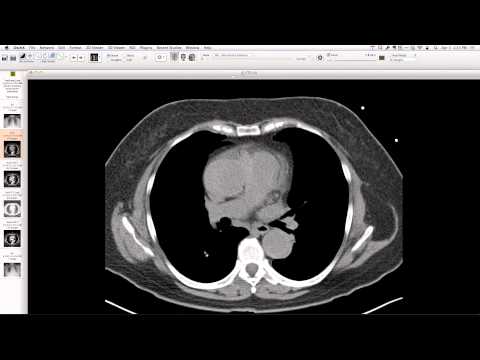
Chest radiograph shows a mildly elongated aorta. Non-contrast CT reveals hyperattenuating intramural hematoma within the thickened wall of the ascending aorta, extending to involve a portion of the pulmonary artery. The contrast-enhanced CT confirms a thickened aortic wall, with subtle out-pouching of contrast from the aortic lumen into the wall, indicating a small communication. These findings were surgically confirmed.
Key takeaways
Acute intramural hematoma (IMH) is a form of acute aortic syndrome. This case demonstrates IMH involving both the ascending aorta and the pulmonary artery, a rare extension. The presence of a small communication between the aortic lumen and the hematoma suggests early progression toward dissection or rupture, emphasizing the importance of both non-contrast and contrast imaging for diagnosis and characterization.
AI-assisted summary — may contain errors. Verify against the source video. Learn more
More from this webinar
- Williams Syndrome with Supravalvular Aortic Stenosis and Pulmonary Artery NarrowingCongenital
- Alveolar Capillary Dysplasia with Unroofed Coronary Sinus (Right-to-Left Shunt)Congenital
- Iatrogenic Esophageal Rupture from Blakemore Tube PlacementIatrogenic
- Primum Atrial Septal Defect (ASD)Congenital
- Pulmonary Alveolar MicrolithiasisMetabolic
- Esophageal Varices Anastomosing with an Inferior Pulmonary VeinVascular
- Unroofed Coronary Sinus (Isolated Finding)Congenital
- Complex Congenital Anomalies: Right Aortic Arch with Aberrant Innominate Artery, Total Anomalous Pulmonary Venous Return of Left Lung, and Cor Triatriatum SinisterCongenital
See all cases from April 3, 2014 →
Related Vascular cases
- Recanalized Pulmonary Arteriovenous Malformation (AVM)
- Liver Enhancement Mimicking Metastasis from SVC Obstruction with Systemic-to-Pulmonary Vein Shunting
- Systemic-to-Pulmonary Vein Shunting from SVC Obstruction in Treated Breast Cancer
- Massive Paraesophageal Varices in End-Stage Liver Disease
- Aneurysmal Aortic Dissection and Rupture in Marfan Syndrome
- Chronic Type B Aortic Dissection with Renal Malperfusion