 CTICases
Bilateral Malpositioned Pleural Drains (Sub-Diaphragmatic and Chest Wall)
 ▶ Watch on YouTube — age-restricted, cannot embed here
[...]. Bilateral Malpositioned Pleural Drains (Sub-Diaphragmatic and Chest Wall). CTICases. Published April 3, 2014. https://cticases.com/c/n4v5a
Imaging findings
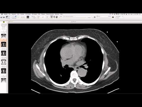
Initial chest radiograph after pleural drain placement shows a left pneumothorax and right upper lobe collapse, with neither pigtail catheter forming a characteristic loop, suggesting malposition. CT confirms that the left-sided drain is located within the chest wall, and the right-sided drain follows a sub-diaphragmatic course, clearly outside the pleural space. Aspiration pneumonitis and right lower lobe collapse are also present.
Key takeaways
This case demonstrates bilateral malposition of pleural drains following emergent placement for a presumed (likely iatrogenic) pneumothorax. The failure of pigtail catheters to loop correctly on chest radiographs is a critical sign of malposition, which in this case included a chest wall placement and a sub-diaphragmatic placement. Prompt CT evaluation is essential to identify such complications and guide correct placement.
AI-assisted summary — may contain errors. Verify against the source video. Learn more
More from this webinar
- Williams Syndrome with Supravalvular Aortic Stenosis and Pulmonary Artery NarrowingCongenital
- Alveolar Capillary Dysplasia with Unroofed Coronary Sinus (Right-to-Left Shunt)Congenital
- Iatrogenic Esophageal Rupture from Blakemore Tube PlacementIatrogenic
- Primum Atrial Septal Defect (ASD)Congenital
- Pulmonary Alveolar MicrolithiasisMetabolic
- Esophageal Varices Anastomosing with an Inferior Pulmonary VeinVascular
- Unroofed Coronary Sinus (Isolated Finding)Congenital
- Complex Congenital Anomalies: Right Aortic Arch with Aberrant Innominate Artery, Total Anomalous Pulmonary Venous Return of Left Lung, and Cor Triatriatum SinisterCongenital
See all cases from April 3, 2014 →
Related Iatrogenic cases
- Dysfunctioning Mechanical Mitral Valve Prosthesis
- Post-Pneumonectomy Bronchopleural Fistula and Empyema
- Anastomotic Bronchial Stenosis after Lung Transplantation
- Right Bronchial Anastomotic Dehiscence after Bilateral Lung Transplant
- Hepatopleural Fistula after Microwave Ablation of Hepatocellular Carcinoma
- Dislodged Vascular Access Port