 CTICases
Diffuse Pulmonary Lymphangiomatosis
 ▶ Watch on YouTube — age-restricted, cannot embed here
[...]. Diffuse Pulmonary Lymphangiomatosis. CTICases. Published July 7, 2014. https://cticases.com/c/q7b8r
Imaging findings
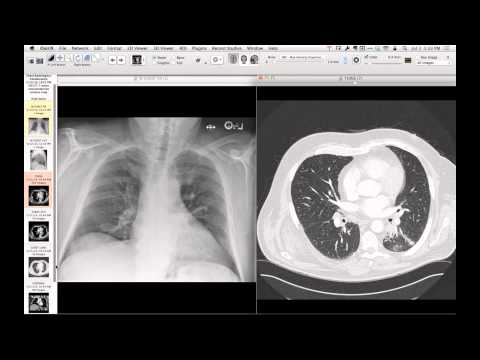
Early lymphangiogram and CT demonstrate contrast in the abdominal lymphatics and supraclavicular nodes, accompanied by large bilateral pleural effusions. Follow-up CT obtained four years later shows severe low-attenuation thickening of the pleura, chest wall, and mediastinal tissues, a pericardial effusion, and diffuse pulmonary lymphatic infiltration characterized by thickened interlobular septa, peribronchovascular bundle thickening, and subpleural lines.
Key takeaways
Diffuse pulmonary lymphangiomatosis is a rare, progressive disease characterized by the abnormal proliferation of lymphatic vessels infiltrating the lungs, mediastinum, pleura, and chest wall. The imaging findings mimic other diffuse lymphatic patterns such as sarcoidosis, Kaposi sarcoma, and lymphangitic carcinomatosis. The clinical course is progressive and can be exacerbated by physiological stressors such as pregnancy.
AI-assisted summary — may contain errors. Verify against the source video. Learn more
More from this webinar
- Spontaneous Pneumomediastinum and Pneumoretroperitoneum due to Macklin PhenomenonOther
- Thoracic Spine Discitis-Osteomyelitis and Paraspinal Abscess due to Disseminated Mycobacterium avium complexInfection
- Right Ventricular Perforation and Pericardial Contrast InjectionIatrogenic
- Esophagopericardial FistulaOther
- Congenital Hypoplasia of the Right Upper LobeCongenital
- Goodpasture Syndrome (Anti-GBM Disease)Autoimmune
- Behçet Syndrome with Multiple Pulmonary Artery AneurysmsAutoimmune
- Left Ventricular Pseudoaneurysm post-TAVRIatrogenic
See all cases from July 7, 2014 →