 CTICases
Fontan Physiology (Contrast Mixing and Streaming Artifacts)
 ▶ Watch on YouTube — age-restricted, cannot embed here
[...]. Fontan Physiology (Contrast Mixing and Streaming Artifacts). CTICases. Published July 17, 2014. https://cticases.com/c/u27g4
Imaging findings
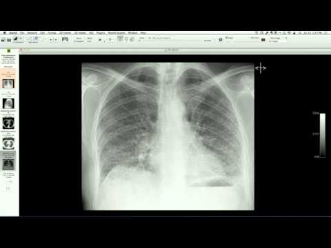
Contrast-enhanced chest CT in a 22-year-old with repaired complex congenital heart disease (single ventricle, single atrium, transposition of the great arteries, and an extracardiac Fontan conduit) shows intense contrast layering and "streaming" artifacts within the pulmonary arteries, creating pseudo-filling defects that mimic pulmonary embolism. Associated mosaic lung attenuation is present due to regional vascular over-perfusion.
Key takeaways
Patients with Fontan physiology rely on passive systemic venous pressure to perfuse the lungs, resulting in slow, non-turbulent blood flow that produces prominent contrast streaming and layering artifacts on CT. This can easily be mistaken for acute pulmonary embolism; recognition of the post-operative congenital anatomy and the unmixed nature of the contrast is crucial to avoid a false-positive diagnosis.
AI-assisted summary — may contain errors. Verify against the source video. Learn more
More from this webinar
- Aortic Arch DissectionVascular
- Sclerosing PeriaortitisInflammatory
- Hypoplastic Left Heart Syndrome (Post-Norwood and Sano Shunt)Congenital
- Tetralogy of Fallot with Ductal Origin of the Left Pulmonary ArteryCongenital
- Migrating Pleural Mesothelial CystCongenital
- Chronic Diaphragmatic RuptureTrauma
- Bronchopulmonary DysplasiaCongenital
- Granulomatous-Lymphocytic Interstitial Lung DiseaseILD
See all cases from July 17, 2014 →
Related Artifact cases
- Bilateral Pleural Effusions Masked on Digital Radiography
- Digital Radiography Artifacts: Grid Misalignment and Backscatter
- Pleural Effusion mimicking Post-Operative Aortic Rupture and Mediastinal Hematoma
- Bifurcated Silicone Tracheal Stent for Tracheomalacia
- Benign Emptying of Post-Pneumonectomy Space
- Leadless Pacemaker (Micra)