 CTICases
Perforation of Right Atrial Pacemaker Lead into Lung
 ▶ Watch on YouTube — age-restricted, cannot embed here
[...]. Perforation of Right Atrial Pacemaker Lead into Lung. CTICases. Published April 3, 2014. https://cticases.com/c/xjd5q
Imaging findings
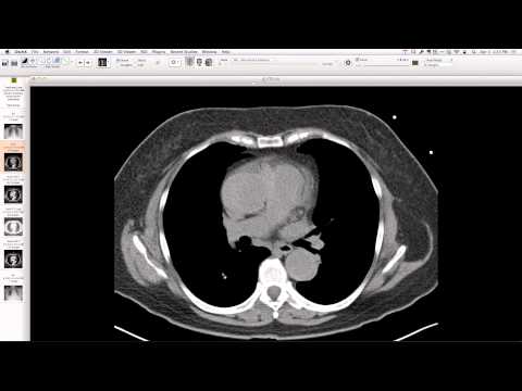
Post-placement radiographs initially showed an apparently well-positioned right atrial pacemaker lead. Subsequent CT demonstrates the right atrial lead perforating the right atrium and extending into the anterior segment of the right upper lobe. No significant scarring around the lead tip was noted.
Key takeaways
Pacemaker lead perforation, while more commonly seen with right ventricular leads, can also occur with right atrial leads. This case highlights a right atrial lead perforating into the lung parenchyma, which can be initially subtle on plain radiographs and may not always be associated with immediate complications like hemothorax or significant tissue damage from electrical pulse, though pneumothorax is a known risk.
AI-assisted summary — may contain errors. Verify against the source video. Learn more
More from this webinar
- Williams Syndrome with Supravalvular Aortic Stenosis and Pulmonary Artery NarrowingCongenital
- Alveolar Capillary Dysplasia with Unroofed Coronary Sinus (Right-to-Left Shunt)Congenital
- Iatrogenic Esophageal Rupture from Blakemore Tube PlacementIatrogenic
- Primum Atrial Septal Defect (ASD)Congenital
- Pulmonary Alveolar MicrolithiasisMetabolic
- Esophageal Varices Anastomosing with an Inferior Pulmonary VeinVascular
- Unroofed Coronary Sinus (Isolated Finding)Congenital
- Complex Congenital Anomalies: Right Aortic Arch with Aberrant Innominate Artery, Total Anomalous Pulmonary Venous Return of Left Lung, and Cor Triatriatum SinisterCongenital
See all cases from April 3, 2014 →
Related Iatrogenic cases
- Dysfunctioning Mechanical Mitral Valve Prosthesis
- Post-Pneumonectomy Bronchopleural Fistula and Empyema
- Anastomotic Bronchial Stenosis after Lung Transplantation
- Right Bronchial Anastomotic Dehiscence after Bilateral Lung Transplant
- Hepatopleural Fistula after Microwave Ablation of Hepatocellular Carcinoma
- Dislodged Vascular Access Port