 CTICases
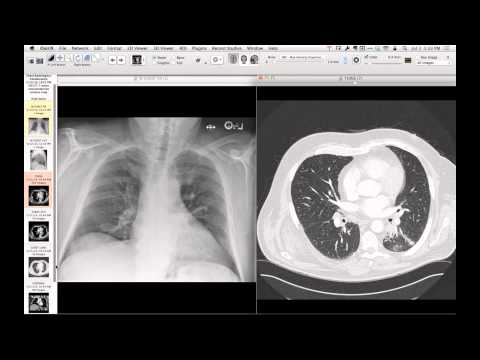
Mediastinal Lipomatosis
 ▶ Watch on YouTube — age-restricted, cannot embed here
[...]. Mediastinal Lipomatosis. CTICases. Published July 7, 2014. https://cticases.com/c/zc3t5
Imaging findings
Chest radiography demonstrates a large anterior mediastinal mass with a positive hilum overlay sign. Chest CT shows a massive, low-attenuation fat-containing mass in the anterior mediastinum causing severe compressive atelectasis of the adjacent lungs, with focus areas of higher-attenuation, complex soft tissue representing necrosis.
Key takeaways
Mediastinal lipomatosis is a benign proliferation of fat that can occasionally mimic a lipomatous tumor. When a fat-dominant mediastinal mass is exceedingly large, causes significant mass effect, or contains thick septations, nodular non-fatty components, or necrosis, histographical evaluation is required to rule out well-differentiated liposarcoma or liposarcomatous transformation.
AI-assisted summary — may contain errors. Verify against the source video. Learn more
More from this webinar
- Spontaneous Pneumomediastinum and Pneumoretroperitoneum due to Macklin PhenomenonOther
- Thoracic Spine Discitis-Osteomyelitis and Paraspinal Abscess due to Disseminated Mycobacterium avium complexInfection
- Right Ventricular Perforation and Pericardial Contrast InjectionIatrogenic
- Esophagopericardial FistulaOther
- Congenital Hypoplasia of the Right Upper LobeCongenital
- Goodpasture Syndrome (Anti-GBM Disease)Autoimmune
- Behçet Syndrome with Multiple Pulmonary Artery AneurysmsAutoimmune
- Left Ventricular Pseudoaneurysm post-TAVRIatrogenic
See all cases from July 7, 2014 →