 CTICases
Chronic Pulmonary Hypertension and Ventricular Septal Defect
 ▶ Watch on YouTube — age-restricted, cannot embed here
[...]. Chronic Pulmonary Hypertension and Ventricular Septal Defect. CTICases. Published July 7, 2014. https://cticases.com/c/28wqz
Imaging findings
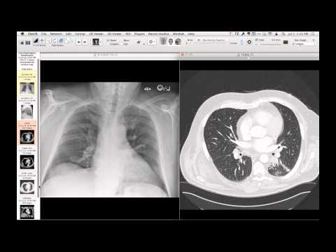
Chest radiography and CT demonstrate massive enlargement of the main, right, and left pulmonary arteries with extensive intimal atherosclerosis and heavily calcified in situ thrombus. An undiagnosed ventricular septal defect is present. The peripheral pulmonary vasculature demonstrates a characteristic corkscrew, tortuous, and beaded neovascular pattern.
Key takeaways
Chronic, uncorrected left-to-right shunts (such as a ventricular septal defect) lead to severe, progressive pulmonary hypertension. Over time, high pressures cause aneurysmal dilation of the central pulmonary arteries, atherosclerosis, and in situ calcified thrombosis, along with peripheral neovascular remodeling.
AI-assisted summary — may contain errors. Verify against the source video. Learn more
More from this webinar
- Spontaneous Pneumomediastinum and Pneumoretroperitoneum due to Macklin PhenomenonOther
- Thoracic Spine Discitis-Osteomyelitis and Paraspinal Abscess due to Disseminated Mycobacterium avium complexInfection
- Right Ventricular Perforation and Pericardial Contrast InjectionIatrogenic
- Esophagopericardial FistulaOther
- Congenital Hypoplasia of the Right Upper LobeCongenital
- Goodpasture Syndrome (Anti-GBM Disease)Autoimmune
- Behçet Syndrome with Multiple Pulmonary Artery AneurysmsAutoimmune
- Left Ventricular Pseudoaneurysm post-TAVRIatrogenic
See all cases from July 7, 2014 →