 CTICases
Spontaneously Resolving Pericardial Diverticulum
 ▶ Watch on YouTube — age-restricted, cannot embed here
[...]. Spontaneously Resolving Pericardial Diverticulum. CTICases. Published July 7, 2014. https://cticases.com/c/2wjug
Imaging findings
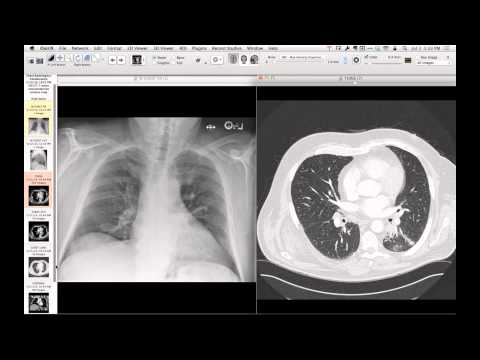
Initial chest CT shows a homogeneous, fluid-attenuation cystic mass in the anterior mediastinum abutting the superior pericardium, suggesting a thymic or pericardial cyst. Follow-up CT demonstrates complete disappearance of the lesion, with only a small residual pericardial diverticulum remaining.
Key takeaways
Pericardial diverticula are congenital, fluid-filled outpouchings that communicate directly with the pericardial space. Unlike static pericardial cysts, diverticula can spontaneously empty and fill due to changes in patient positioning, intrathoracic pressure, or pericardial fluid volume, resulting in their transient disappearance on follow-up imaging.
AI-assisted summary — may contain errors. Verify against the source video. Learn more
More from this webinar
- Spontaneous Pneumomediastinum and Pneumoretroperitoneum due to Macklin PhenomenonOther
- Thoracic Spine Discitis-Osteomyelitis and Paraspinal Abscess due to Disseminated Mycobacterium avium complexInfection
- Right Ventricular Perforation and Pericardial Contrast InjectionIatrogenic
- Esophagopericardial FistulaOther
- Congenital Hypoplasia of the Right Upper LobeCongenital
- Goodpasture Syndrome (Anti-GBM Disease)Autoimmune
- Behçet Syndrome with Multiple Pulmonary Artery AneurysmsAutoimmune
- Left Ventricular Pseudoaneurysm post-TAVRIatrogenic
See all cases from July 7, 2014 →