 CTICases
Acute Hypersensitivity Pneumonitis (Gray Lungs/Black Bronchus Sign)
 ▶ Watch on YouTube — age-restricted, cannot embed here
[...]. Acute Hypersensitivity Pneumonitis (Gray Lungs/Black Bronchus Sign). CTICases. Published March 4, 2016. https://cticases.com/c/3bcp8
Imaging findings
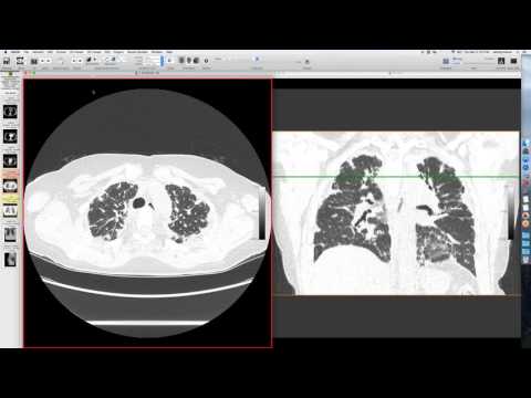
A patient with restrictive lung disease and DLCO abnormalities had a chest radiograph and CT showing diffuse bilateral 'gray lungs,' characterized by extensive and confluent central lobular nodules. Some air trapping was also noted.
Key takeaways
This is a classic presentation of hypersensitivity pneumonitis, demonstrating the 'gray lungs' appearance and the 'black bronchus sign,' where the blackness of air in the bronchi stands out against the diffuse ground-glass opacification of the lungs. Follow-up imaging showed significant improvement after treatment. Hypersensitivity pneumonitis is a form of ILD.
AI-assisted summary — may contain errors. Verify against the source video. Learn more
More from this webinar
- Fat Embolism SyndromeTrauma
- Dystrophic Calcification from Rhabdomyolysis and Pulmonary Interstitial EmphysemaMetabolic
- Nocardiosis in a Heart Transplant PatientInfection
- Pleuroparenchymal Fibroelastosis as a Manifestation of Graft-versus-Host DiseaseILD
- Synchronous Lung Cancer and Pulmonary HamartomaNeoplastic
- Variant Coronary Artery in the Ligament of MarshallCongenital
- Job's Syndrome (Hyper-IgE Syndrome) with Chronic Staphylococcal InfectionsAutoimmune
- Squamous Cell Carcinoma Obstructing Bronchus IntermediusNeoplastic
See all cases from March 4, 2016 →
Related ILD cases
- Chronic Hypersensitivity Pneumonitis (Bird Fancier's Lung)
- Chronic Hypersensitivity Pneumonitis (Pigeon Breeder's Lung) with Air Trapping
- Smoking-Related Bronchiolitis/DIP Mimicking Hypersensitivity Pneumonitis
- Progressive Langerhans Cell Histiocytosis (PLCH)
- Lupus with Cystic Lung Destruction (mimicking honeycombing)
- Bleomycin-induced Lung Injury