 CTICases
Multiple Traumatic Aortic Tears
 ▶ Watch on YouTube — age-restricted, cannot embed here
[...]. Multiple Traumatic Aortic Tears. CTICases. Published July 17, 2014. https://cticases.com/c/4v64t
Imaging findings
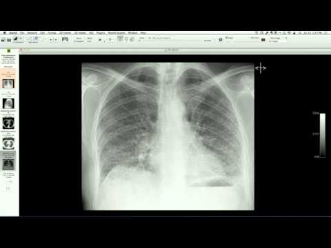
Chest radiograph shows mediastinal widening, tracheal deviation, a left apical cap, and a left hemithorax pleural effusion. Contrast-enhanced CT angiography demonstrates a severe multi-segment traumatic aortic injury with tears in the proximal (isthmus), mid, and lower descending thoracic aorta, accompanied by an extensive mediastinal hematoma tracking into the anterior mediastinum and between the aorta and trachea.
Key takeaways
Severe blunt chest trauma (such as high-speed motor vehicle collisions) can cause multiple simultaneous tears along the thoracic aorta, rather than a single focal tear at the classic aortic isthmus. These high-energy injuries present with profound mediastinal hematoma and typically require emergency endovascular repair with multi-segment stent grafts.
AI-assisted summary — may contain errors. Verify against the source video. Learn more
More from this webinar
- Aortic Arch DissectionVascular
- Sclerosing PeriaortitisInflammatory
- Hypoplastic Left Heart Syndrome (Post-Norwood and Sano Shunt)Congenital
- Tetralogy of Fallot with Ductal Origin of the Left Pulmonary ArteryCongenital
- Migrating Pleural Mesothelial CystCongenital
- Chronic Diaphragmatic RuptureTrauma
- Bronchopulmonary DysplasiaCongenital
- Granulomatous-Lymphocytic Interstitial Lung DiseaseILD
See all cases from July 17, 2014 →