 CTICases
Empyema following Chest Trauma
 ▶ Watch on YouTube — age-restricted, cannot embed here
[...]. Empyema following Chest Trauma. CTICases. Published August 20, 2015. https://cticases.com/c/5j9my
Imaging findings
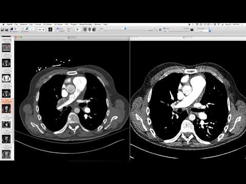
Frontal and lateral radiographs showed a very large, non-effacing opacity in the left hemithorax, consistent with a posterior pleural space process. The lateral projection showed a large lenticular shape of an intrapleural fluid collection with gas/air within it. CT confirmed a large pleural fluid collection with air, consistent with empyema. A couple of rib fractures were noted from a fall weeks prior.
Key takeaways
Empyema, especially after chest trauma, can present as a large, lenticular, intrapleural fluid collection with air. Radiographic signs like non-effacement of vessels on frontal view and a lenticular shape on lateral view are key to distinguishing it from intraparenchymal lesions. Elderly patients, sometimes with impaired immunity, can develop extensive empyema with minimal symptoms.
AI-assisted summary — may contain errors. Verify against the source video. Learn more
More from this webinar
- Retained Wires from First-Generation DefibrillatorIatrogenic
- Hyperacute Pulmonary Infarction progressing to Acute PE with Pleural EffusionVascular
- Left Upper Lobe Anomalous Pulmonary Venous Stricture (Incidental)Congenital
- Lymphocytic Interstitial Pneumonia (LIP) in association with B-cell LymphomaNeoplastic
- Endobronchial Papillomatosis with Left Lower Lobe CollapseNeoplastic
- Esophageal Carcinoma with Radiographic CorrelationNeoplastic
- Cardiogenic Pulmonary Edema with Ground Glass OpacityVascular
- Hypersensitivity Pneumonitis (HP) in a Dog GroomerILD
See all cases from August 20, 2015 →
Related Infection cases
- Mycoplasma-induced Acute Interstitial Pneumonia / ARDS
- Disseminated Mycobacterium Bovis (BCG) Infection Presenting as Pleural and Chest Wall Disease
- E. coli Sternal Osteomyelitis and Anterior Mediastinitis
- Hematogenous Dissemination of Enteric Organisms Causing Costal Chondritis and Chest Wall Cellulitis
- Legionella Pneumonia
- Blastomycosis