 CTICases
Lymphangioleiomyomatosis presenting as Spontaneous Pneumothorax
 ▶ Watch on YouTube — age-restricted, cannot embed here
[...]. Lymphangioleiomyomatosis presenting as Spontaneous Pneumothorax. CTICases. Published October 30, 2015. https://cticases.com/c/fatu9
Imaging findings
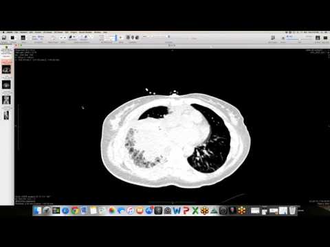
Initial chest radiograph shows a large unilateral pneumothorax with underlying coarse, diffuse reticular opacities. Subsequent chest CT obtained after pleural catheter placement reveals extensive, thin-walled, round bilateral lung cysts of uniform size distributed symmetrically throughout all lung zones.
Key takeaways
Lymphangioleiomyomatosis is a progressive cystic lung disease that almost exclusively affects women of childbearing age, often presenting acutely with a spontaneous pneumothorax. On chest radiographs, the diffuse cysts can easily be mistaken for a reticular pattern, but chest CT is highly specific, showing uniform, thin-walled, round cysts with normal intervening lung parenchyma.
AI-assisted summary — may contain errors. Verify against the source video. Learn more
More from this webinar
- Beta-Thalassemia with Extramedullary Hematopoiesis and Diffuse Marrow ExpansionMetabolic
- Anomalous High Origin of the Right Coronary Artery with Incidental Developing PneumoniaCongenital
- Pulmonary Hemorrhage Secondary to Acute Pulmonary Embolism presenting as Abdominal PainVascular
- Acute Exacerbation of Idiopathic Pulmonary Fibrosis Triggered by Minor TraumaILD
- Type A Intramural Hematoma with Focal Intimal TearVascular
- Aortic Rupture and Mediastinal Hematoma Decompressing into the Right LungVascular
- Congenital Left Main Coronary Artery Osteal AtresiaCongenital
- Pleural Extramedullary Hematopoiesis in MyelofibrosisMetabolic
See all cases from October 30, 2015 →
Related Neoplastic cases
- Post-Transplant Lymphoproliferative Disorder (PTLD), Polyclonal Polymorphic Type
- Primary Effusion Lymphoma (PFL) in HIV-Positive Patient
- Multifocal Adenocarcinoma with Tracheal Invasion
- Metastatic Breast Cancer Mimicking Primary Lung Cancer
- Pulmonary Carcinoid Tumor (mimicking DIPNECH)
- Metastatic Prostate Cancer Invading Trachea and Mediastinum