 CTICases
Pulmonary Hemorrhage Secondary to Acute Pulmonary Embolism presenting as Abdominal Pain
 ▶ Watch on YouTube — age-restricted, cannot embed here
[...]. Pulmonary Hemorrhage Secondary to Acute Pulmonary Embolism presenting as Abdominal Pain. CTICases. Published October 30, 2015. https://cticases.com/c/y53r3
Imaging findings
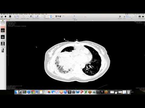
Abdominal CT extending through the lung bases and dedicated non-contrast chest CT demonstrate extensive ground-glass opacity, interstitial and bronchial wall thickening, and confluent consolidation in the right lower lobe, accompanied by a pleural effusion. Contrast-enhanced CT reveals a large, occlusive pulmonary embolus within the right lower lobe pulmonary artery.
Key takeaways
Lower lobe pulmonary embolism can cause diaphragmatic irritation and refer pain to the abdomen, prompting initial evaluation via abdominal CT. Pulmonary hemorrhage without true alveolar necrosis is pathologically far more common than true pulmonary infarction following an embolism. On CT, these infarctions or hemorrhages classically present as peripheral consolidations lacking air bronchograms, which can easily be misdiagnosed as routine bacterial pneumonia.
AI-assisted summary — may contain errors. Verify against the source video. Learn more
More from this webinar
- Beta-Thalassemia with Extramedullary Hematopoiesis and Diffuse Marrow ExpansionMetabolic
- Anomalous High Origin of the Right Coronary Artery with Incidental Developing PneumoniaCongenital
- Acute Exacerbation of Idiopathic Pulmonary Fibrosis Triggered by Minor TraumaILD
- Type A Intramural Hematoma with Focal Intimal TearVascular
- Aortic Rupture and Mediastinal Hematoma Decompressing into the Right LungVascular
- Congenital Left Main Coronary Artery Osteal AtresiaCongenital
- Pleural Extramedullary Hematopoiesis in MyelofibrosisMetabolic
- Mediastinal Gray Zone LymphomaNeoplastic
See all cases from October 30, 2015 →
Related Vascular cases
- Double Saddle Pulmonary Embolism
- Interstitial Lung Edema with Fluid in Interlobar Fissures
- Chronic Pulmonary Thromboembolism (CTEPH)
- Reverse Takotsubo Cardiomyopathy
- Spontaneous Auto-Embolization of a Pulmonary Arteriovenous Malformation (AVM)
- Type A Aortic Dissection with Acute Bilateral Lower Extremity Paralysis and Retroperitoneal Hematoma