 CTICases
Aortic Rupture and Mediastinal Hematoma Decompressing into the Right Lung
 ▶ Watch on YouTube — age-restricted, cannot embed here
[...]. Aortic Rupture and Mediastinal Hematoma Decompressing into the Right Lung. CTICases. Published October 30, 2015. https://cticases.com/c/6nvp2
Imaging findings
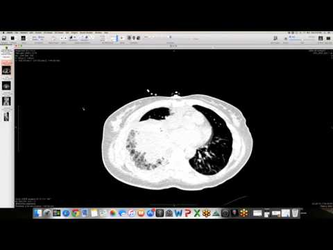
Serial CT studies show a focal area of ascending aortic wall calcification that progresses over years to develop localized outpouchings and pseudoaneurysms. This eventually culminates in a frank, contained rupture of the ascending aorta, presenting as a large mediastinal hematoma that decompresses directly into the right upper and middle lobes of the lung.
Key takeaways
Atherosclerotic plaque and calcification in the ascending aorta can progress to form penetrating atherosclerotic ulcers or pseudoaneurysms. If these lesions undergo rupture under high arterial pressure, they can decompress directly into the adjacent lung parenchyma, mimicking focal consolidative pneumonia or pulmonary hemorrhage on imaging.
AI-assisted summary — may contain errors. Verify against the source video. Learn more
More from this webinar
- Beta-Thalassemia with Extramedullary Hematopoiesis and Diffuse Marrow ExpansionMetabolic
- Anomalous High Origin of the Right Coronary Artery with Incidental Developing PneumoniaCongenital
- Pulmonary Hemorrhage Secondary to Acute Pulmonary Embolism presenting as Abdominal PainVascular
- Acute Exacerbation of Idiopathic Pulmonary Fibrosis Triggered by Minor TraumaILD
- Type A Intramural Hematoma with Focal Intimal TearVascular
- Congenital Left Main Coronary Artery Osteal AtresiaCongenital
- Pleural Extramedullary Hematopoiesis in MyelofibrosisMetabolic
- Mediastinal Gray Zone LymphomaNeoplastic
See all cases from October 30, 2015 →
Related Vascular cases
- Double Saddle Pulmonary Embolism
- Interstitial Lung Edema with Fluid in Interlobar Fissures
- Chronic Pulmonary Thromboembolism (CTEPH)
- Reverse Takotsubo Cardiomyopathy
- Spontaneous Auto-Embolization of a Pulmonary Arteriovenous Malformation (AVM)
- Type A Aortic Dissection with Acute Bilateral Lower Extremity Paralysis and Retroperitoneal Hematoma