 CTICases
Type A Intramural Hematoma with Focal Intimal Tear
 ▶ Watch on YouTube — age-restricted, cannot embed here
[...]. Type A Intramural Hematoma with Focal Intimal Tear. CTICases. Published October 30, 2015. https://cticases.com/c/3exra
Imaging findings
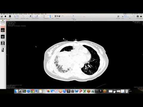
Non-contrast CT demonstrates a high-attenuation crescent within the wall of the ascending aorta consistent with an intramural hematoma, accompanied by hemopericardium and mediastinal soft-tissue stranding. Contrast-enhanced CT reveals a focal, subtle intimal defect and a linear filling defect measuring approximately 1.5 cm along the medial aspect of the ascending aorta just above the aortic root.
Key takeaways
An intramural hematoma associated with a localized, short-segment intimal tear is sometimes referred to as a limited aortic dissection. Because transthoracic and transesophageal echocardiograms can frequently be false-negatives in the setting of limited intimal defects, high-resolution electrocardiogram-gated CT is crucial to detect the entry tear and guide emergent surgical repair for type A lesions.
AI-assisted summary — may contain errors. Verify against the source video. Learn more
More from this webinar
- Beta-Thalassemia with Extramedullary Hematopoiesis and Diffuse Marrow ExpansionMetabolic
- Anomalous High Origin of the Right Coronary Artery with Incidental Developing PneumoniaCongenital
- Pulmonary Hemorrhage Secondary to Acute Pulmonary Embolism presenting as Abdominal PainVascular
- Acute Exacerbation of Idiopathic Pulmonary Fibrosis Triggered by Minor TraumaILD
- Aortic Rupture and Mediastinal Hematoma Decompressing into the Right LungVascular
- Congenital Left Main Coronary Artery Osteal AtresiaCongenital
- Pleural Extramedullary Hematopoiesis in MyelofibrosisMetabolic
- Mediastinal Gray Zone LymphomaNeoplastic
See all cases from October 30, 2015 →
Related Vascular cases
- Double Saddle Pulmonary Embolism
- Interstitial Lung Edema with Fluid in Interlobar Fissures
- Chronic Pulmonary Thromboembolism (CTEPH)
- Reverse Takotsubo Cardiomyopathy
- Spontaneous Auto-Embolization of a Pulmonary Arteriovenous Malformation (AVM)
- Type A Aortic Dissection with Acute Bilateral Lower Extremity Paralysis and Retroperitoneal Hematoma