 CTICases
Beta-Thalassemia with Extramedullary Hematopoiesis and Diffuse Marrow Expansion
 ▶ Watch on YouTube — age-restricted, cannot embed here
[...]. Beta-Thalassemia with Extramedullary Hematopoiesis and Diffuse Marrow Expansion. CTICases. Published October 30, 2015. https://cticases.com/c/e9k4w
Imaging findings
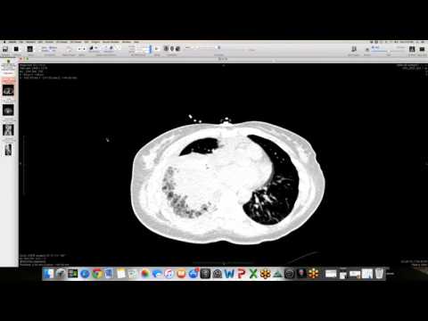
Coronal CT and MR images demonstrate striking, diffuse expansion of the medullary spaces of all visualized ribs. Multiple soft-tissue density, iso-intense to muscle paraspinal masses are present along the posterior mediastinum in the paravertebral regions. These masses exhibit heterogeneous enhancement on post-contrast series. Additionally, MR imaging demonstrates that the liver is extremely dark on T2-weighted and gradient echo sequences.
Key takeaways
Diffuse marrow expansion and extramedullary hematopoiesis are classic compensatory responses to chronic severe anemia in patients with beta-thalassemia. Extramedullary hematopoiesis classically manifests as soft-tissue paraspinal masses in the posterior mediastinum. Due to repeated blood transfusions, these patients often develop secondary hemochromatosis, resulting in marked iron deposition and T2-shortening (darkening) of the liver on MRI.
AI-assisted summary — may contain errors. Verify against the source video. Learn more
More from this webinar
- Anomalous High Origin of the Right Coronary Artery with Incidental Developing PneumoniaCongenital
- Pulmonary Hemorrhage Secondary to Acute Pulmonary Embolism presenting as Abdominal PainVascular
- Acute Exacerbation of Idiopathic Pulmonary Fibrosis Triggered by Minor TraumaILD
- Type A Intramural Hematoma with Focal Intimal TearVascular
- Aortic Rupture and Mediastinal Hematoma Decompressing into the Right LungVascular
- Congenital Left Main Coronary Artery Osteal AtresiaCongenital
- Pleural Extramedullary Hematopoiesis in MyelofibrosisMetabolic
- Mediastinal Gray Zone LymphomaNeoplastic
See all cases from October 30, 2015 →