 CTICases
Progressive Coccidioidomycosis Cavity
 ▶ Watch on YouTube — age-restricted, cannot embed here
[...]. Progressive Coccidioidomycosis Cavity. CTICases. Published March 4, 2016. https://cticases.com/c/qmf64
Imaging findings
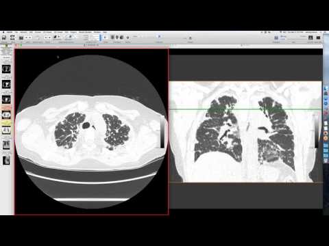
A patient with known coccidioidomycosis had a small cavity in the left upper lobe in 2011. Over three years, it progressively enlarged, initially with a smooth wall, then becoming thicker, more irregular, and inflammatory, possibly communicating with the airway. By 2015 and 2016, the cavity continued to grow, showing some fluid and calcium along its wall.
Key takeaways
While coccidioidomycosis cavities typically resolve in about 85% of patients, this case demonstrates a chronic, progressive course over four years that did not regress, eventually requiring surgical resection. Hesitation to operate on coccidioidomycosis lesions exists due to the risk of contaminating the pleural space and spreading the infection. Additionally, strict precautions are necessary when handling specimens to prevent laboratory personnel from contracting the disease.
AI-assisted summary — may contain errors. Verify against the source video. Learn more
More from this webinar
- Fat Embolism SyndromeTrauma
- Dystrophic Calcification from Rhabdomyolysis and Pulmonary Interstitial EmphysemaMetabolic
- Nocardiosis in a Heart Transplant PatientInfection
- Pleuroparenchymal Fibroelastosis as a Manifestation of Graft-versus-Host DiseaseILD
- Synchronous Lung Cancer and Pulmonary HamartomaNeoplastic
- Variant Coronary Artery in the Ligament of MarshallCongenital
- Job's Syndrome (Hyper-IgE Syndrome) with Chronic Staphylococcal InfectionsAutoimmune
- Squamous Cell Carcinoma Obstructing Bronchus IntermediusNeoplastic
See all cases from March 4, 2016 →
Related Infection cases
- Coccidioidomycosis Presenting as Lobar Pneumonia in a Renal Transplant Patient
- Achalasia Complicated by Mycobacterium fortuitum Pneumonia
- Coxsackie Viral Constrictive Pericarditis
- Pulmonary Coccidioidomycosis Mimicking Metastasis
- Recurrent Infective Endocarditis with Mycotic Pulmonary Aneurysms
- Discitis-Osteomyelitis