 CTICases
Idiopathic Pulmonary Fibrosis
 ▶ Watch on YouTube — age-restricted, cannot embed here
[...]. Idiopathic Pulmonary Fibrosis. CTICases. Published July 17, 2014. https://cticases.com/c/s6mss
Imaging findings
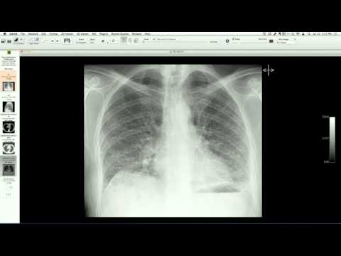
Chest radiograph and CT demonstrate subpleural, basilar-predominant reticular opacities and traction bronchiectasis without macroscopic honeycombing. Surgical lung biopsy confirms histopathological features of usual interstitial pneumonia (UIP), including fibroblast foci and temporal heterogeneity.
Key takeaways
Under current diagnostic guidelines, a CT pattern showing basilar-predominant subpleural reticulation and traction bronchiectasis without honeycombing is classified as "possible UIP" or "probable UIP". However, in the appropriate clinical context, the majority of these cases demonstrate a classic UIP pattern on histopathology, confirming idiopathic pulmonary fibrosis (IPF).
AI-assisted summary — may contain errors. Verify against the source video. Learn more
More from this webinar
- Aortic Arch DissectionVascular
- Sclerosing PeriaortitisInflammatory
- Hypoplastic Left Heart Syndrome (Post-Norwood and Sano Shunt)Congenital
- Tetralogy of Fallot with Ductal Origin of the Left Pulmonary ArteryCongenital
- Migrating Pleural Mesothelial CystCongenital
- Chronic Diaphragmatic RuptureTrauma
- Bronchopulmonary DysplasiaCongenital
- Granulomatous-Lymphocytic Interstitial Lung DiseaseILD
See all cases from July 17, 2014 →